Tag Archives: Rehabilitation Services

How to get relief from muscle strain injury?

Treatment of Quadriceps Muscle Strain Injury (Front Thigh Muscle Pain):

Introduction

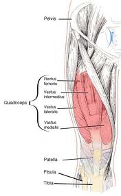
Strains of the quadriceps muscle usually occur during sprinting, jumping or kicking. Strains are seen in all the quadriceps muscles but are most common in the rectus femoris, which is more vulnerable to strain as it passes over two joints: the hip and the knee. The most common site of strain is the distal musculotendinous junction of the rectus femoris. Management of this type of rectus femoris strain and of strains of the vast muscles is relatively straightforward; rehabilitation time is short. Strains of the proximal rectus are not as straightforward and considered separately below.

Types of Quadriceps Muscle Strain

  • Mild (Grade 1)
  • Moderate (Grade II)
  • Severe (Grade III)

Like all muscle strains, quadriceps strains may be graded into mild (grade 1), moderate (grade II) or severe, complete tears (grade III). The athlete feels the injury as a sudden pain in the anterior thigh during an activity requiring explosive muscle contraction.

There is local pain and tenderness and, if the strain is severe, swelling and bruising. Grade I strain is a minor injury with pain on resisted active contraction and on passive stretching. An area of local spasm is palpable at the site of pain. An athlete with such a strain may not cease activity at the time of the pain but will usually notice the injury after cooling down or the following day.

Moderate or grade II strains cause significant pain on passive stretching as well as on unopposed active contraction. There is usually a moderate area of inflammation surrounding a tender palpable lesion. The athlete with a grade II strain is generally unable to continue the activity. Complete tears of the rectus femoris occur with sudden onset of pain and disability during intense activity. A muscle fiber defect is usually palpable when the muscle is contracted. In the long term, they resolve with conservative management, often with surprisingly little disability.

Treatment of Quadriceps Muscle Strain

The principles of treatment of a quadriceps muscle strain are similar to those of a thigh contusion.  They are also appropriate for the treatment of quadriceps strain; however, depending on the severity of the strain, progression through the various stages may be slower.

  • Although loss of range of motion may be less obvious than with a contusion, it is important that the athlete regain pain-free range of movement as soon as possible.
  • Loss of strength may be more marked than with a thigh contusion and strength retraining requires emphasis in the rehabilitation program.
  • As with the general principles of muscle rehabilitation, the program should commence with low resistance, high repetition exercise.
  • Concentric and eccentric exercises should begin with very low weights.
  • General fitness can be maintained by activities such as swimming (initially with a pool buoy) and upper body training.
  • Functional retraining should be incorporated as soon as possible.
  • Full training must be completed prior to return to sport. Unfortunately, quadriceps strains often recur, either in the same season, or even a year to two later.

Differentiating between a Mild Quadriceps Strain and a Quadriceps Contusion

Occasionally, it may be difficult between a minor contusion and a minor muscle strain but the distinction needs to be made as an athlete with a thigh strain should progress more slowly through a rehabilitation program than should the athlete with quadriceps contusion. The athlete with thigh strain should avoid sharp acceleration and deceleration movements in the early stages of injury. Some of the features that may assist the clinician in differentiating. Diagnostic ultrasound examination may be helpful in differentiating between the two conditions.

DIAGNOSTIC FEATURES QUADRICEPS CONTUSION
Mechanism Contact Injury
Pain Onset Immediate or soon after
Location Usually Lateral or Distal
Bruising/Swelling May be obvious early
Effect of gentle stretch May initially aggravate pain
Strength testing No loss of strength except pain inhibition.
Behavior of pain Improves with gentle activity.


Active Physical Therapy provides best services .Our services include to physical therapy, occupational therapy, hand therapy, senior wellness, neurological rehabilitation, orthopedic rehabilitation, industrial rehabilitation and specialties including auto accident injuries / Trauma Cases, Work-related Injuries, Sports Injuries, Post Surgical Rehabilitation, Lower Back Pain, Shoulder Pain/ Injuries, , Arthritis, Bursitis/Tendonitis, Carpal Tunnel Syndrome, Knee and Ankle Injuries, Leg Pain, Lumbar Stabilization, Muscle Strains. We severe many locations. For More Information Call Now at : 301-498-1604

http://www.active-physicaltherapy.com/

Tendonitis of Ankle and Foot Pain

Treatment of  Flexor Hallucis Longus Tendinopathy:

The flexor hallucis longus tendon flexes the big toe and assists in plantarflexion of the ankle. It passes posterior to the medial malleolus, and runs between the two sesamoid bones to insert into the base of the distal phalanx of the big toe.

Causes:

  • Flexor hallucis longus tendinopathy may occur secondary to overuse, a stenosing tenosynovitis, pseuocyst or tendon tear.
  • A common cause is overuse in a ballet dancer, as dancers repetitively go from flat foot stance to the en pointe position, when extreme plantarflexion is required. Wearing shoes that are too big and require the athlete to ‘toe-grip’ may also result in flexor hallucis longus tendinopathy.
  • This condition is often associated with posterior impingement syndrome as the flexor hallucis tendon lies in a fibro-osseous tunnel between the lateral and medial tubercles of the posterior process of the talus.
  • Enlargement or medial displacement of the os trigonum puts pressure on the flexor hallucis longus at the point where the tendon changes direction from a vertical course dorsal to the talus to a horizontal course beneath the talus.
  • This can cause tendon thickening and may result in ‘triggering’ of the tendon, when partial tearing and subsequent healing of the tendon produce excessive scar tissue.

Clinical Features:

  • Pain on toe-off or forefoot weight-bearing (e.g. rising in ballet), maximal over the posteromedial aspect of the calcaneus around the sustentaculum tali.
  • Pain may be aggravated by resisted flexion of the first toe or stretch into full dorsiflexion of the hallux.
  • In more severe cases, there may be ‘triggering’ of the first toe, both with rising onto the balls of the foot (e.g. in ballet) and in lowering from this position. Triggering occurs when the foot is placed in plantarflexion and the athlete, unable to flex the hallux, but then with forcible active contraction of the flexor hallucis longus, is able to extend the interphalangeal or metatarsophalangeal joints of the toe. A snap or pop occurs in the posteromedial aspect of the ankle when this happens. Subsequent passive flexion or extension of the interphalangeal joint produces a painless snap posterior to the medial malleolus.

Diagnosis of Flexor Hallucis Longus Tendinopathy:

MRI or ultrasound may both reveal pathology. The characteristic MRI sign is abrupt fluid cut-off in the tendon sheath; excessive fluid is found loculated around a normal-appearing tendon proximal to the fibro-osseous canal.

Treatment of Flexor Hallucis Longus Tendinopathy:

In the acute phase, treatment may include:

  • Ice
  • Avoidance of activities that stress the flexor hallucis longus tendon (e.g. dancer working at the barre but not rising en pointe)
  • Flexor hallucis longus strength and stretching exercises
  • Soft tissue therapy proximally in the muscle belly
  • Correction of subtalar joint hypomobility with manual mobilization
  • Control of excessive pronation during toe-off with tape or orthoses-this may be helpful but is difficult to achieve in dancers.

Prevention of  Flexor Hallucis Longus Tendinopathy:

Prevention of recurrences should focus on a reduction in the amount of hip turnout, thus ensuring that the weight is directly over the hip, avoidance of hard floors, and using firm, well-fitting pointe shoes, so that the foot is well supported and no additional strain is placed on the tendon. Technique correction is important in ballet dancers with this condition as it is thought to arise not only from excessive ankle eversion or inversion with pointe work but also from proximal weakness, such as poor trunk control. Surgical  should be considered when persistent synovitis or triggering prevents dancing en pointe. Surgery involves exploration of the tendon and release of the tendon sheath.

Gastrocnemius Muscle Strains ( Calf Pain)

Symptoms and Treatment of Acute Calf Strain:

Acute strain of the gastrocnemius muscle occurs typically when the athlete attempts to accelerate from a stationary position with the ankle in dorsiflexion, or when lunging forward, such as while playing tennis or squash. Sudden eccentric overstretch, such as when an athlete runs onto a kerb and the ankle drops suddenly into dorsiflexion, is another common mechanism.

The exact moment of injury was caught on video in the case of a famous Australian batsman whose gastrocnemius strain occurred when has entire body weight was over his foot on the injured side with the center of mass well in front of the leg. The gastrocnemius muscle-tendon complex was at close to maximal length, and the muscle-tendon length was also constant at the time. Therefore, the injury probably occurred just as the muscle-tendon complex was moving from an eccentric to an isometric phase.

Signs and Symptoms

  • The patient complains of an acute, stabbing or tearing sensation usually either in the medial belly of the gastrocnemius or at the musculotendinous junction.
  • Examination reveals tenderness at the site of muscle strain.
  • Stretching the gastrocnemius reproduces pain, as doe’s resisted plantar flexion with the knee extended.
  • In grade III muscle tears, there may be a palpable defect.
  • Assess functional competence of the injured muscle by asking the patient to perform a bilateral heel raise.
  • If necessary, a unilateral heel raise, a heel drop or hop may be used to reproduce the pain.
  • This places the muscle under progressively greater load concentrically and eccentrically.
  • Calf muscle strain can be graded.
  • The tightness of the muscle itself should be assessed as overuse may often lead to palpable ropelike bands or local tissue thickening, which may predispose to further injury.

Treatment

Initial treatment aims to reduce pain and swelling with the use of ice and electrotherapeutic modalities (e.g. TENS, magnetic field therapy, interferential stimulation).

  • Crutches may be necessary if the patient is unable to bear weight.
  • A heel raise should be used on both the injured and uninjured side.
  • Gentle stretching of the gastrocnemius to the level of a feeling of tightness can begin soon after injury.
  • Muscle strengthening should start after 24 hours.
  • This involves a progression of exercises, commencing with concentric bilateral calf raise, followed by unilateral calf raise with the gradual addition of weights and, finally, eccentric calf lowering over a step gradually increasing speed, then adding weights.
  • Low-impact cross-training such as stationary cycling or swimming can be commenced as soon as pain allows.
  • When active weight-bearing muscle contraction is pain-free, sustained myofascial tension should be performed on the muscle belly with digital ischemic pressure to focal areas of increased tone and/or tenderness.
  • Endeavor to correct possible predisposing factors, such as calf muscle tightness, that may arise from poor biomechanics.
  • Athletes should undergo a graduated return to weight-bearing, progressing through walking, easy jogs and, as eccentric strength returns, include sprint and change of direction drills.

Tennis leg

  • The term tennis leg refers to an acute muscle tear in the older athlete characterized by sudden onset of severe calf pain and significant disability.
  • The injury is invariably associated with extensive bruising and swelling, and can be mistaken for a deep venous thrombosis.
  • The most common site is the medial head of gastrocnemius, but occasionally the planters muscle is involved.

Acute Nerve Root Compression

Symptoms And Treatment of Herniated Disc:

Acute nerve root compression is usually the result of an acute disk prolapse when the contents of the nucleus pulposus of the intervertebral disk are extruded through a defect in the anulus fibrosus into the spinal canal. There they may irritate the nerve root. The irritation of the nerve root may be due to direct mechanical compression by the nuclear material or as result of the chemical irritation caused by the extrusion. In the older athlete, nerve roots may be compressed by osteophytes formed as part of a degenerative process.

Prolapse usually occurs in disks that have been previously damaged by one of the processes mentioned already. This explains why frequently a minor movement, such as bending over to pick up an object off the floor, may cause such an apparently severe injury. Disk prolapse usually occurs between the ages of 20 and 50 years and is more common in males than females. The L5-S1 disk is the most commonly propalsed disk and L-4-5 the next most common.

Signs and Symptoms

Typically, a patient with a disk prolapse presents with acute low back pain or radicular leg pain (or both) following a relatively trivial movement usually involving flexion.

  • On occasions, the presentation may be painless, with weakness or sensory symptoms only.
  • The symptoms depend on the direction of the extrusion.
  • Posterior protrusions are more likely to cause low back pain with later development of leg pain, whereas posterolateral protrusions may cause radicular symptoms without low back pain.
  • Typical symptoms include sharp shooting pain in a narrow band accompanied by pins and needles, numbness and weakness.
  • Pain is often aggravated by sitting, bending, lifting, coughing or sneezing.
  • Pain is usually eased by lying down, particularly on the asymptomatic and is often less after a night’s rest.
  • On examination, the patient often demonstrates a list to one side, usually, although not always, away from the side of pain.
  • This is a protective scoliosis. Examination may be difficult if there is severe pain and irritability.
  • Straight leg raise is usually limited (less than 30 degree in severe cases) and all active movements, particularly flexion, are usually restricted.
  • Palpation usually reveals acute muscle spasm with marked tenderness but occasionally it may be unremarkable.
  • A neurological examination should always be performed when pain extends past the buttock fold or there are subjective sensory/motor changes.

Treatment

In the acute phase, the most appropriate treatment is rest in bed in a position of maximum comfort with administration of analgesics and NSAIDs.

  • The patient should lie as much as possible and avoid sitting.
  • Extension exercises should be commenced as soon as possible. However, if exercises cause an increase in peripheral symptoms, they should be ceased.
  • Mobilization techniques should be performed with great care.
  • Rotations may be effective but should be performed gently as patients with disk prolapse may be made considerably worse with aggressive mobilization.
  • Manipulation is contraindicated in conditions with acute neurological signs and symptoms.
  • Traction is often helpful in the treatment of acute disk prolapsed with distal symptoms.
  • However, it is not uncommon for the patient to experience considerable pain relief while undergoing traction, only to have increased symptoms after treatment.
  • A transforaminal epidural injection of corticosteroid may help if there is no significant improvement in symptoms and signs with rest.
  • Surgery may be required if neurological signs persist or worsen. If bowel or bladder symptoms are present, emergency surgery may be necessary.
  • An open Laminectomy or percutaneous diskectomy using a needle aspiration technique may be performed.
  • Chymopa pain injection may be helpful when a unilateral disk bulge is present.
  • As the acute episode settles, it is important to restore normal pain-free movement to the area with localized mobilization and stretching.

 Following restoration of range of movement, active stabilization exercises should be performed. Postural advice, including correction of poor lifting techniques and adjustment of sporting technique, where necessary, is most important.

Shoulder Instability

Symptoms And Treatment of Anterior Instability:

Shoulder Instability may be anterior, posterior, inferior or multidirectional.

Anterior Instability

Anterior glenohumeral instability may be post-traumatic, as a result of an acute episode of trauma causing anterior dislocation or subluxation, or a traumatic, or a combination-for instance, an acute traumatic episode in a lax shoulder.

In differentiating between the two types of anterior instability, the history is the most useful factor. In post-traumatic instability, the patient usually reports a specific incident that precipitated the problem. This is commonly a moderately forceful abduction and external rotation injury. Following this episode, however, the patient reports that the shoulder has never returned to normal. In many post-traumatic types of instability a true dislocation may not have occurred and the symptoms are related to recurrent subluxation. The atraumatic type of abnormality is common in people with capsular laxity including sportspeople, especially those involved in repeated overhead activities such as baseball pitchers, javelin throwers, swimmers and tennis players.

Symptoms of Anterior Instability

The symptoms of anterior instability include recurrent dislocation or subluxation, shoulder pain and episodes of dead arm syndrome.

  • Pain usually arises from impingement of the rotator cuff tendons with recurrent anterior translation of the humeral head and recurrent silent subluxation.
  • This is aggravated by the eventual weakening of the rotator cuff muscles which, in turn, fail to depress the humeral head adequately.
  • The recurrent episodes of impingement result in a rotator cuff tendinopathy.
  • Anterior shoulder pain in association with post-traumatic anterior instability may be due to catching of a labral detachment.
  • This pain and sensation of catching may be reproduced on anterior drawer or load and shift testing.
  • The dead arm effect is thought to arise from traction or impingement on the neurovascular structures, causing transient numbness and weakness of the arm.
  • This usually resolves after a few minutes. The episodes of subluxation and dislocation usually increase in frequency.

Occasionally, a stage is reached where relatively minor activities such as yawning or rolling over in bed may result in a subluxation or dislocation.

Treatment

As outlined earlier, a traditional sling should not be used to manage instability.

  • If aggressive non-operative treatment is to be pursued, then the arm should be placed in external rotation of 30 degree for three weeks night and day to reduce the Bankart lesion.
  • There are a large number of different procedures used to treat shoulder instability. In athletes, particularly those whose dominant throwing arm is involved, the underlying mechanical lesion should be corrected.
  • In most cases, this involves repair of the Bankart lesion, which may be performed either as an open or arthroscopic procedure.
  • Other mechanical problems such as a tear in the rotator cuff may also be corrected.
  • If an extremely large Hill’-Sachs lesion is present, then a procedure such as bone grafting may be necessary.
  • In treating atraumatic instability, intensive rehabilitation involves strengthening of the dynamic stabilizers (rotator cuff muscles) and scapular stabilizing muscles, with particular emphasis on the muscles opposing the direction of the instability.
  • Modification of sporting activity may also be helpful. If conservative measures fail, then surgery should be considered .This usually involves a capsular shift procedure.

Cardiovascular Fitness

Sports Activities with Rehab Treatment:

The maintenance of cardiovascular fitness is another essential component of the rehabilitation process. No matter what type of injury the athlete has sustained, it should always be possible to design an exercise program to enable cardiovascular fitness to be maintained.

In injuries to the lower limb that require a period of restricted weight-bearing activity, cardiovascular fitness may be maintained by performing activities such as cycling, swimming or water exercises. These activities can be used in a training program that follows the same principles as the athlete’s normal training. Depending on the athlete’s particular sport, this may include a combination of endurance, interval, anaerobic and power work.

It is important to maintain these alternative training methods for cardiovascular fitness even after the patient has resumed some weight-bearing training of his or her own. The clinician must explain to the patient that while he or she is gradually returning to weight-bearing activity, the cardiovascular endurance aspect of training should be performed as non-weight-bearing.

Following complete recovery and return to sport, it may be advantageous, particularly in patients who have had an overuse injury, to incorporate some of these non-weight-bearing forms of training as a substitute for some weight-bearing training.

Progression of Rehabilitation

There are several different parameters that the therapist may manipulate to progress the athletes program to a level at which return to sport is possible. These parameters are:

  • Type of Activity
  • Duration of Activity
  • Frequency of Activity/Rest
  • Intensity of Activity
  • Complexity of Activity

Type of Activity

In the early stages of the rehabilitation program, we recommend activities that do not directly stress the injured area. However, these exercises may still result in some mobilization or strengthening of the injured area, for example, tennis ground strokes following an ankle injury, cycling following shoulder impingement. Later in the program, activities specifically involving the injured area will test its integrity and prepare it for functional activity.

Duration of Activity

Once the activity is directly stressing the injured area, the time spent performing that activity must be increased very gradually. It is advisable to slowly increase the amount of time spent performing a particular activity, for example, jogging, and then hold it constant at a particular level and vary one of the other parameters, such as frequency.

Frequency

An integral part of the rehabilitation program is recovery. It allows tissues to adapt to the stress of exercise. For example, a runner with Achilles tendinopathy may initially run every third day, then every second day, then two out of every three days and ultimately six or seven days per week. On non-running days, the athlete should maintain fitness by swimming or cycling as well as performing the other elements of the rehabilitation program, for example, muscle strengthening.

Intensity

As the athlete progresses through the rehabilitation program, the intensity (speed and power) of the activity will increase. A rehabilitation program for a sprinter may involve progression from half pace to three-quarter pace to full pace. Race starts will be included later in the program. Other variables include surfaces and shoes. Progression can be made from softer surfaces to harder surfaces or from flat running shoes to spikes once full speed is achieved.

Complexity of Activity

The athlete can progress from simple activities to more complex movements. For example, a basket-baller dribbles slowly in a straight line gradually increasing speed and introducing turns, or a tennis player progresses from ground strokes to incorporating overhead shots and rallying drills before playing points competitively and ultimately six or seven days per week. On non-running days, the athlete should maintain fitness by swimming or cycling as well as performing the other elements of the rehabilitation program, for example, muscle strengthening.

Exercise Prescription in Patients with Osteoporosis

Benefits And Tips of  Exercises Activities:

Most types of activity programs are preferable to a sedentary lifestyle for patients with osteoporosis and with a 20-minute warm-up, comprising 10 minutes of gentle stretching and range of motion activity followed by 10 minutes of aerobic exercises. For cardiovascular exercises, targeted heart rate should be 60% of maximum heart rate (220 – age) for a beginner or a deconditioned woman and 70-75% for those in more intermediate health. Weight training with light free weights and rubber tubing can then be incorporated.

Benefits for Osteoporosis Exercise

As per standard resistance training, the exercises should target the major muscle groups.

  • Effective upper arm exercises include pushing against a wall or pulling and twisting against a partner.
  • Quadriceps strength can be improved with a wall slide (squat) exercise or by practicing standing from a seated position.
  • Trunk stabilization (i.e. core stability) exercises are often introduced in the crooklying position and progressed to sitting and standing.
  • Trunk stabilization exercises target the recruitment of transversus abdominis and the internal and external obliques rather than rectus abdominis.
  • Due to the propensity to develop a kyphotic posture with osteoporosis, back posture correction exercises should also be emphasized and can be done standing, in a chair or prone.
  • Balance exercises can be introduced initially by having individuals hold a tandem stance for 10 seconds with their eyes open while holding onto the kitchen counter for support.
  • Other balance exercises include single leg stance with eyes open (then eyes closed), tandem forward walking, walking backwards, and tandem backwards walking.
  • The exercise program should conclude with a 15-minute cool down period and warm-down stretches can be done sitting on the floor.
  • Remember also that the assessment for exercise prescription should allow the practitioner to discover what activities the client values.
  • Many popular activities require some degree of strength, flexibility, endurance, balance and coordination. For example, a good line dancing class emphasizes posture and the attributes listed, is fun, and does not require a partner.

Community Based Exercise Program For Osteoporosis

‘Osteofit’ is an exercise-based program devised by staff of the British Columbia Women Health Center Osteoporosis Program in Vancouver, Canada. This community-based program for women and men aims to reduce participant’s risk of falling and improve their functional ability and thereby enhance their quality of life. It differs from typical seniors exercise classes by specifically targeting posture, balance, gait, coordination, and trunk and pelvic stabilization rather than general aerobic fitness.

‘Osteofit Tips’ For Exercise

A typical class consists of a warm-up, the workout and a relaxation component, which are outlined below. ‘Osteofit’ classes also include ‘Osteofit Tips’ a 5-minute health education topic that the instructor shares with participants.

  • The work-out itself consists of strengthening and stretching exercises intended to improve posture by combating medially rotated shoulders, chin protrusion (excessive cervical extension), thoracic kyphosis and loss of lumbar lordosis.
  • Exercises to improve balance and coordination may progress from heel raises and toe pulls to the mildly challenging two-legged heel-toe rock and the more challenging tandem walks and obstacle courses.
  • Pelvic stabilization is trained using leg exercises (e.g. hip abduction and extension) or balance exercises.
  • After appropriate training and progression through less challenging positions, trunk stabilization is addressed when the participant is cued and positioned to do all standing exercises with resistance for the arms (e.g. biceps curls) and shoulders (e.g. lateral arm raises).
  • The abdominal muscles are strengthened in their function as stabilizers rather than as prime movers.
  • Exercises to improve functional ability include chair squats and getting up and down off the floor.
  • Upper and lower body activities are alternated to reduce the risk of tendinopathy.
  • If the class includes more than one set of an exercise, the sets are separated by a short rest period.
  • Repetitions are kept to between eight and 16 and weights are relatively light so that participants do not work to fatigue with each set.
  • The exercises are arranged so that the less strenuous exercises, such as hamstring stretching, are at the end of the work-out.
  • The last few minutes of the class are devoted to relaxation techniques such as deep breathing, progressive muscle tensing and relaxing, and visualizations to a background of soft music and/or nature sounds.
  • ‘Osteofit‘ is one form of safe and effective exercise for a population that is at high risk of osteoporotic fracture.

Treatment of Injuries

Sports Injuries,Acute Injuries And Overuse Injuries:

Regular physical activity is probably the most important overall determinant of a population’s health. Unfortunately, physical activity may extract a cost in the form of an activity-related injury. Such an injury may be categorised as either being an acute injury or an overuse injury depending on the mechanism of injury and the onset of symptoms.

Acute Injuries

Acute injuries may be due to extrinsic causes, such as a direct blow, either as a result of contact with another player or equipment, or intrinsic causes, such as a ligament sprain or muscle tear. Acute injuries may be classified according to the particular site injured (e.g. bone, cartilage, joint,ligament, muscle, tendon, bursa, nerve or skin) and the type of injury (e.g. fracture, dislocation, sprain or strain).

Overuse Injuries

Overuse injuries present three distinct challenges to the clinician-diagnosis, treatment and an understanding of why the injury occurred. Diagnosis requires taking a comprehensive history of the onset, nature and site of the pain along with a thorough assessment of potential risk factors, for example, training and technique. Careful examination may reveal which anatomical structure is affected. It is often helpful to ask patients to perform the maneuver that produces their pain.

Causes of Overuse Injuries

A cause must be sought for every overuse injury. The cause may be quite evident,

  • Such as a sudden doubling of training quantity,
  • Poor footwear or an obvious biomechanical abnormality or may be more subtle, such as running on a cambered surface, muscle imbalance or leg length discrepancy.
  • The causes of overuse injuries are usually divided into extrinsic factors such as training, surfaces, shoes, equipment and environmental conditions.
  • Intrinsic factors such as malalignment, leg length discrepancy, muscle imbalance, muscle weakness, lack of flexibility and body composition.
  • Possible factors in the development of overuse injuries.

Treatment of Overuse Injuries

The treatment of overuse injuries may include relative rest, that is,

  • Avoidance of aggravating activities
  • While maintaining fitness
  • The use of ice and various electrotherapeutic modalities
  • Soft tissue techniques; and drugs
  • Such as the non-steroidal anti-inflammatory drugs (NSAIDs).

Bone

Stress fractures

Stress fractures, a common injury among sportspeople, were first reported in military recruits in the l9th century. A stress fracture is a micro fracture in bone that results from repetitive physical loading below the single cycle failure threshold. Overload stress can be applied to bone through two mechanisms:

  • The redistribution of impact forces resulting in increased stress at focal points in bone.
  • The action of muscle pulls across bone.

Histological changes resulting from bone stress occur along a continuum beginning with vascular congestion and thrombosis. This is followed by osteoclastic and osteoblastic activity leading to rarefaction, weakened trabeculae and micro fracture and ending in complete fracture. This sequence of events can be interrupted at any point in the continuum if the process is recognized.

Similarly, the process of bony remodeling and stress fracture in athletes is recognized as occurring along clinical continuum with pain or radiographic changes presenting identifiable markers along the continuum. Since radioisotopic imaging and MRI can detect changes in bone at the phase of accelerated remodeling, these investigations can show stress-induced bony changes early in the continuum.

Stress fractures may occur in virtually any bone in the body. The most commonly affected bones are the tibia, metatarsals, fibula, tarsal navicular, femur and pelvis. A list of sites of stress fractures and the likely associated sports or activities.

It is important to note that a bone scan, although a routine investigation for stress fractures, is non-specific, and other bony abnormalities such as tumors and osteomyelitis may cause similar pictures. It may also be difficult to localize the site of the area of increased uptake precisely,especially in an area such as the foot where numerous small bones are in close proximity.

Diagnostic Features of A Stress Fracture

  • Localized pain and tenderness over the fracture site.
  • A recent change in training or taking up a new activity.
  • X-Ray appearance is often normal or there may be a periosteal reaction.
  • Abnormal appearance on radioisotopic bone scan scintingraphy), CT scan or MRI.

MRI is being increasingly advocated as the investigation of choice for stress fractures. Even though MRI does not image fractures as clearly as do computed tomography (CT) scans, it is of comparable sensitivity to radioistopic bone scans in assessing bony damage. The typical MRI appearance of a stress fracture shows periosteal and marrow edema plus or minus the actual fracture line.

Treatment of Stress Fractures

The treatment of stress fractures generally requires avoidance of the precipitating activity.

  • The majority of stress fractures heal within six weeks of beginning relative rest.
  • Healing is assessed clinically by the absence of local tenderness and functionally by the ability to perform the precipitating activity without pain.
  • It is not useful to attempt to monitor healing with X-ray or radioisotopic bone scan.
  • CT scan appearances of healing stress fractures can be deceptive as in some cases the fracture is still visible well after clinical healing has occurred.
  • The return to sport after clinical healing of a stress fracture should be a gradual process to enable the bone to adapt to an increased load.
  • An essential component of the management of an overuse injury is identification and modification of risk factors.
  • There are, however, a number of sites of stress fractures in which delayed union or non-union of the fracture commonly occurs.
  • These fractures need to be treated more aggressively.
  • The sites of these fractures and the recommended treatments.

Bone Strain

In some athletes bone scans show uptake of radioisotope at non-painful sites. This is thought to represent bony remodeling at a very early subclinical level and has been termed bone strain. Another situation encountered in clinical practice is the painful tender focal area of bone that demonstrates a mildly increased uptake of radioisotope on bone, insufficient to be classified as a stress fracture. This has been termed ‘stress reaction’. It would appear that there is a continuum of bone response to stress that ranges from mild (bone strain) to severe (stress fracture). The clinical features of bone strain, stress reaction and stress fractures.

The presence of bone strain or a stress reaction are probably an indication that the patient is moving further along the continuum towards a stress fracture and should probably be an indication for reduction or modification of activity.

Articular cartilage

Overuse injury can affect the articular cartilage lining of joints, particularly in osteoarthritis. Changes range from microscopic inflammatory changes to softening, fibrillation, fissuring and ultimately to gross visible changes. In younger people, this pathology can arise at the patella (patellofemoral syndrome), but it is important to note that the pain of patellofemoral syndrome can occur in the presence of normal joint surfaces.

Joint

Inflammatory changes in joints associated with over use are classified as synovitis or capsulitis. Examples of these problems are the sinus tarsi syndrome of the subtalar joint and synovitis of the hip joint.

Ligament

Overuse injuries of ligaments are uncommon. The medial collateral ligament of the knee occasionally becomes inflamed, particularly in breaststroke swimmers.

Muscle

Focal tissue thickening/fibrosis

Repetitive microtrauma caused by overuse damages muscle fibers. This is thought by some to lead to development of adhesions between muscle fibers and formation of cross-linkages in fascia.

Clinically, these changes may be palpated as firm focal areas of tissue thickening, taut, thickened bands arranged in the direction of the stress or as large areas of increased muscle tone and thickening.

These lesions may cause local pain or predispose other structures, such as tendons, to injury due to reduction in the ability of the tissue to elongate under stretch or eccentric load. This will also compromise the ability of the affected muscle to contract an relax rapidly.

These minor muscle injuries, which occur frequently in association with hard training, may respond to regular soft tissue therapy, strengthening and stretching.

Tendon

Tendon injuries are among the most common overuse injuries. Tendons, which are made up of tight parallel collagen bundles, transmit forces from muscle to bone and are, therefore, subject to great tensile stresses. Tendons withstand strong tensile forces, resist shear forces less well and provide little resistance to compression force. The stress-strain curve for tendons. As the strain increases, tissue deformation begins, some fibers begin to fall and ultimately macroscopic tendon failure occurs. There is, however, a large margin between the stresses that cause tendon failure and those that are experienced during normal physiological loading.

The vasculature of tendons is variable with the blood supply originating at both the musculotendinons and bone-tendon junctions. Vascular tendons are surrounded by a paratenon and avascular tendons are in sheaths. Tendon vascularity is compromised at site of friction, torsion or compression.

Bursa

The body contains many bursae situated usually between bony surfaces and overlying tendons. Their role is to facilitate movement of the tendon over the bony surface. Overuse injuries in bursae are quite common, particularly at the subacromial bursa, the greater trochanteric bursa, the bursa deep to the iliotibial band at the knee and the retrocalcaneal bursa separating the Achilles tendon from the calcaneus.

Bursitis is associated with local tenderness and swelling and pain on specific movements. Treatment involves the use of NSAIDs but this may be ineffective due to the poor blood supply of most bursae. Occasionally, a bursa needs to be drained of its fluid with or without subsequent corticosteroid infiltration.

Nerve

Nerve entrapment syndromes occur in athletes as a result of swelling in the surrounding soft tissues or anatomical abnormalities. These may affect the suprascapular nerve, the posterior interosseous, ulnar and median nerves in the forearm, the obturator nerve in the groin, the posterior tibial nerve at the tarsal tunnel on the medial aspect of the ankle and, most commonly, the interdigital nerves, especially between the third and fourth toes, a condition known as a Morton’s neuroma. This condition is not a true neuroma but rather a nerve compression. These nerve entrapments occasionally require surgical decompression.

Chronic Mild irritation of a nerve may result in damage manifested by an increase in neural tension. These may be the primary cause of the patient’s symptoms or may contribute to symptoms.

Skin

Blisters may occur at any site of friction with an external source or sporting equipment. Foot blisters can be prevented by wearing-in new shoes, wearing socks , and smearing petroleum jelly over the sock at sites of friction .Strategies to prevent blisters also serve to prevent callus. symptomistic callus can be pared down with a scalpel blade, taking care not to lacerate the normal skin.

At the first sign of a blister, the aggravating source should be removed and either adhesive tape applied over the blistered area or blister pads should be applied. Blister pads prevent blisters by acting as a barrier between skin and shoe. Treatment of blisters involves prevention of infection by the use of antiseptics and protection with sticking plaster. Fluid-filled blisters may be punctured and drained.

Clinical Presentation of Patient’s with Overuse Pain

  • Pain some time after exercise or, more frequently, the following morning upon rising.
  • Can be painful at rest and initially becomes less painful with use.
  • Athletes can run through the pain or the pain disappears when they warm up, only to return after exercise when they cool down.
  • The athlete is able to continue to train fully in the early stages of the condition; this may interfere with the healing process.
  • Examination, local tenderness and thickening.
  • Frank swelling and crepitus may be present, although crepitus is more usually a sign of associated tenosynovitis or is due to the hydrophilic (water attracting)nature of the collagen disarray (it is not ‘inflammatory fluid’).
Joint-Related Symptoms Without Acute Injury

Symptoms of Joint Pain:

The dictum not everything that presents to the sports clinic is sports medicine should never be forgotten. In daily practise sports clinicians see many patients who have mechanical joint injuries; thus, it can be tempting to attribute a mechanical diagnosis to every patient who presents with a painful or swollen joint. It is, however, wise to maintain an index of suspicion for inflammatory joint disease masquerading as a mechanical condition. For example, a 3O-year-old runner may present with recurrent knee swelling but have no convincing history of injury. Swelling is very uncommon in patellofemoral pain and a meniscal injury is rare without trauma at that age. Thus, the athlete presents with a single swollen joint but no injury. The clinician should be alert to the possibility that this swollen knee may be caused by an inflammatory condition.

The diagnosing patients with the following four common presentations:

  • The single swollen  joint
  • Low back pain and stiffness
  • Multiple joint symptoms
  • Joint pain and pain all over

We also discuss when to use rheumatological investigations and how to interpret them.

The Patient with a Single Swollen Joint

In the athlete with a single swollen joint without a history of trauma, a possible inflammatory cause should be considered.  Summarizes the differential diagnosis of a single swollen joint.

Clinical perspective

The key to accurate diagnosis of a swollen joint is through taking a careful history and physical examination and having an appropriate index of suspicion. Inflammatory joint problems are characterized by pain, swelling, warmth, redness, night pain and prominent morning stiffness. In all athletes, and especially in children and adolescents, inflammatory, infective or neoplastic conditions should be considered in the light of these symptoms.

History

Many of the inflammatory diseases are associated with extra-articular features that may provide additional clues as to the diagnosis:

  • Psoriatic arthritis may be associated with rash, nail dystrophy, tendon insertion pain (enthesopathy) or low back pain.
  • A history of inflammatory bowel disease (ulcerative colitis, Crohn’s disease or celiac disease) suggests enteropathic arthritis. Urethral discharge or eye inflammation may suggest a reactive arthritis.
  • Rheumatoid arthritis is characteristically a small joint (hands, wrists and feet), symmetrical polyarthritis but can present as a single swollen joint in 15% of cases.
  • Hypothyroidism, hyperparathyroidism and hemochromatosis may be associated with calcium pyrophosphate dihydrate deposition in articular tissues that may manifest as an acute gout-like presentation (pseudo gout) or may have a sub acute or chronic course.
  • Previous renal disease or diuretic use may give clues to diagnosing gout.
  • Septic arthritis is uncommon in the normal joint but the possibility should be considered in  joints recently aspirated or in patients with arthritis, diabetes or impaired immune function.
  • A family history of inflammatory arthritis is significant as first-degree relatives of patients with rheumatoid arthritis are four times more likely to develop the condition than the general population.

The Patient with Low Back Pain and Stiffness

A systematic illness is present in upto 10% of patients who present with low back pain. Because patients with low back pain gravitate to sports clinicians for management, it is important that clinicians have an index of suspicion for those patients with a non-mechanical cause for their low back pain.

Clinical Perspective

The differential diagnosis of low back pain is broad and is documented. This differential diagnosis includes inflammatory arthritis of the spine and sacroiliac joints, known as spondyloarthropathy. Spondyloarthropathy is a generic term applied to the clinical, radiological and immunological features shared by the following diseases:

  • Ankylosing Spondylitis
  • Reactive Arthritis following genitourinary or gut infection
  • Psoriatic Arthritis
  • Enteropathic Arthritis (Crohns disease, ulcerative colitis or celiac disease).

Although patients with these conditions have an increased likelihood of being positive for HLA B27, a negative result does not eliminate the diagnosis. Spondyloarthropathy has its greatest prevalence in young men and usually achieves near full disease expression by age 35 years; thus, patients commonly present to the sports clinician.

History

Patients with back pain due to spondyloarthropathy complain of pain that is worse at night, with prominent morning stiffness (of 2 hours or more), which is eased with gentle exercise and NSAIDs. This pain pattern is very different from the typical pain pattern of mechanical low back pain. Buttock or posterior thigh pain may be present, so this symptom does not distinguish the two types of back pain. When the patient describes morning back pain with prominent stiffness, the physician should ask whether there is a past history of psoriasis or nail dystrophy (psoriatic arthritis), inflammatory bowel disease (enteropathic arthritis), or recent genitourinary or gut infection (reactive arthritis). Spondyloarthropathy is characterized by inflammation of the entheses, commonly at the patellar tendon, Achilles tendon and the plantar fascia.

Peripheral joints may be involved with spondyloarthropathy, particularly an asymmetric, lower limb, large joint inflammation. The shoulder or hip is involved in 30% of patients with ankylosing  spondylitis. A history of extra-articular involvement such as anterior uveitis (iritis) and the rash of keratoderma blennorrhagica or circinate balanitis (reactive arthritis) may provide clues to the specific cause of back pain. It is important that the clinician actively seeks these associations as the athlete may not find them noteworthy to mention. There is often a strong family history of spondyloarthropathy ; for example, approximately 6% of siblings of patients with ankylosing spondylitis will develop the condition.

The Patient Presenting with Multiple Painful Joints

Occasionally patients may attend the sports medicine clinic with multiple joint pain (polyarthralgia) or multiple joint pain with synovitis (polyarthralgia). A systematic approach is vital to make an accurate diagnosis .Summarizes the differential diagnosis of the patient presenting with a polyarthritis.

History

The practitioner should begin by distinguishing polyarthritis with joint pain, stiffness and swelling from polyarthralgia alone. Joint inflammation is characterized by night pain, prominent morning stiffness (of at least 60 minutes but often for hours), swelling, warmth and loss of function. In many of these conditions the diagnosis is clinical. A key diagnostic feature is the onset and pattern of joint involvement.

Rheumatoid arthritis symmetrically affects the small joints of the hands, wrists and feet (PIP, MCP, MTP) and in the majority of patient’s onset occurs over weeks or months. Reactive arthritis (following genitourinary or gastrointestinal infection), on the other hand, is often more rapid in onset and has a propensity to asymmetric involvement of the large joints of the lower limb together with enthesitis (inflammation at the insertions of tendons, ligaments or capsules) or dactylitis (sausage digits) .The duration of symptoms should be recorded.

Parvovirus B19 polyarthritis frequently affects young women who care for small children (mothers or school teachers) who develop parvovirus B19 infection (fifth disease or slapped cheek syndrome). This condition may be indistinguishable from early rheumatoid arthritis. Symptoms and signs usually settle within six weeks, whereas rheumatoid arthritis often follows a chronic and progressive course. The presence or absence of extra-articular manifestations of rheumatological conditions may also aid accurate diagnosis. The pattern of joint involvement in polyarticular pseudogout or psoriatic arthritis often resembles rheumatoid arthritis but without nodulosis, vasculitis or other systemic features seen in rheumatoid arthritis.

The Patient with Joint Pain who ‘Hurts All Over’

A challenging presentation for any clinician is the evaluation of the athlete with widespread joint or muscle pain who hurts all over. These patients are often frequent attenders and it can be extremely rewarding to provide a diagnosis and the help they need. In many cases, patients with this presentation have little to find on clinical examination .The differential diagnosis of this presentations is broad and includes the conditions listed. Other possible diagnosis and directs the practitioner towards appropriate clinical evaluation.

Investigations must be directed towards a specific diagnosis but may include blood count, ESR, plasma immunoglobulin assay band electrophoresis, calcium, phosphate, thyroid function and creatine kinase tests.

ELBOW PAIN

Lateral Elbow Pain: Causes, Symptoms And Treatment

Use of the upper limb in sport demands a well functioning elbow. In addition, injuries in this region may interfere with the patient’s everyday activities. The clinical approach to elbow pain is considered under the following headings:

  • Lateral Elbow Pain, with a particular focus on
  • Extensor Tendinopathy
  • Medial Elbow Pain
  • Posterior Elbow Pain
  • Acute Elbow Injuries
  • Forearm Pain
  • Upper Arm Pain.

Lateral Elbow Pain

Lateral elbow pain is an extremely common presentation among sportspeople and manual workers. The most common cause is an overuse syndrome related to excessive wrist extension. This condition has traditionally been known as ‘tennis elbow‘. This is an unsatisfactory term as it gives little indication of the pathological processes involved. In fact, the condition is more common in non-tennis players than in tennis players. It has also been referred to as ‘lateral epicondylitis’. This is also inappropriate as the site of the abnormality is usually just below the lateral epicondyle and the primary pathology is due to collagen disarray rather than inflammation.

The primary pathological process involved in this condition is tendinosis of the extensor carpiradials brevis (ECRB) tendon, usually within 1-2 cm of its attachment to the common extensor origin at the lateral epicondyle. This condition will be referred to as extensor tendinopathy.

Other conditions that may cause lateral elbow pain include synovitis of the radio humeral joint, radiohumeralbursitis and entrapment of the posterior interosseous branch of the radial nerve (radial tunnel syndrome). These conditions may exist by themselves or in conjunction with extensor tendinopathy.

There is often a contribution to lateral elbow pain from the cervical and upper thoracic spines and neural structures. This may be a relatively minor contribution or, in some cases, the main cause of the patients elbow pain. A full assessment of the cervical spine and neural structures is essential in examination of the patient with lateral elbow pain.

History

The characteristics of the patients lateral elbow pain should be elicited. The diffuse pain of extensor tendinopathy typically radiates from the lateral epicondyle into the proximal forearm extensor muscle mass. Occasionally the pain may be more localized. The onset of pain may be either acute or insidious. There may have been recent changes in training or technique, note-taking or equipment used in sport or work.

The severity of pain ranges from relatively trivial pain to an almost incapacitating pain that may keep the patient awake at night. It is important to note whether the pain is aggravated by relatively minor everyday activities, such as picking up a cup, or whether it requires repeated activity, such as playing tennis or bricklaying, to become painful.

Pain may radiate into the lateral aspect of the forearm. This may be consistent with posterior interosseous nerve entrapment or irritation of other neural structures. If pain is closely related to the activity level, it is more likely to be of a mechanical origin. If pain is persistent, unpredictable or related to posture, referred pain should be considered.

Certain movements, usually those involving wrist extension or gripping, will aggravate mechanical pain. Referred pain is affected by prolonged posture, such as lengthy periods seated at a desk or in a car. Associated sensory symptoms, such as pins and needles, may indicate a neural component. Presence of neck, upper thoracic or shoulder pain should also be noted.

Often by the time the patient presents to the sports medicine clinician, he or she will already have undergone a variety of treatments. It is important to note the response to each of these treatments.

An activity history should also be taken, noting any recent change in the level of activity. In tennis players, note any change in racquet size, grip size or string tension and whether or not any comment has been made regarding his or her technique.

Examination

Examination involves:

1. Observation from the front

2. Active movements

  • elbow flexion/extension
  • supination/pronation
  • wrist flexion (forearm pronated)
  • wrist extension

3. Passive movements

  • as above

4. Resisted movements

  • wrist extension
  • extension at the third metacarpophalangaI-joint
  • grip test

5. Palpation

  • lateral epicondyle
  • extensor muscles

6. Special tests

  • neural tension
  • cervical spine examination
  • thoracic spine examination
  • periscapular soft tissues

Investigations

Investigations are usually not performed in the straightforward case of lateral elbow pain. However, in longstanding cases, plain X-ray (AP and lateral views) of the elbow may show osteochondritis dissecans, degenerative joint changes or evidence of heterotopic calcification.

Ultrasound examination may prove to be a useful diagnostic tool in the investigation of patients with lateral elbow pain. Ultrasound may demonstrate the degree of tendon damage as well as the presence of a bursa.

Extensor tendinopathy

For this major sports medicine condition, we review the pathology, outline the clinical presentation, and then discuss evidence based and clinically founded treatment.

Clinical Features

Extensor tendinopathy occurs in association with any activity involving repeated wrist extension against resistance. This includes sporting activities, such as tennis ,squash and badminton, as well as occupational and leisure activities, such as carpentry, bricklaying, sewing and knitting. Computer use has been shown to be associated with the development of this condition. The peak incidence is between the ages of 40 and 50 years but this condition may affect any age group.

There are two distinct clinical presentations of this condition. The most common is an insidious onset of pain, which occurs 24-72 hours after unaccustomed activity involving repeated wrist extension. This occurs typically after a person spends the weekend laying bricks or using a screwdriver. It is also seen after prolonged sewing or knitting .In the tennis player, it may occur after the use of a new racquet, playing with wet, heavy balls or over hitting, especially hitting into the wind. It also occurs when the player is hitting ‘late’(getting the position slowly), so that body weight is not transferred correctly and the player relies on the forearm muscles exclusively for power.

Treatment

No single treatment has proven to be totally effective in the treatment of this condition. A combination of the different treatments mentioned below will result in resolution of the symptoms in nearly all cases.

The basic principles of treatment of soft tissues injuries apply. There must be control of pain, encouragement of the healing process, restoration of flexibility and strength, treatment of associated factors (e.g. increased neural tension, referred pain),gradual return to activity with added support and correction of the predisposing factors.

Control of Pain

It remains unclear as to how much pain is ideal in the treatment of tendinopathies. Clinical experience suggests that a low level of pain, which does not worsen with training, is likely to not be harmful for tendon healing. However, some patients require relative rest, application of ice and analgesia for comfort.