The elbow is a hinge joint between the lower end of the humerus bone in the upper arm and the upper end of the radius and ulnar bones in the lower arm. The arm is bent and rotated at the elbow by the biceps muscles in the upper arm. Ligaments located at the front, back, and sides of the elbow help stabilize the joint.
Elbow pain can be caused by many problems. A common cause in adults is tendinitis. This is inflammation and injury to the tendons, which are soft tissues that attach muscle to bone.
Lateral Elbow Pain: Causes, Symptoms And Treatment
Use of the upper limb in sport demands a well functioning elbow. In addition, injuries in this region may interfere with the patient’s everyday activities. The clinical approach to elbow pain is considered under the following headings:
- Lateral Elbow Pain, with a particular focus on
- Extensor Tendinopathy
- Medial Elbow Pain
- Posterior Elbow Pain
- Acute Elbow Injuries
- Forearm Pain
- Upper Arm Pain.
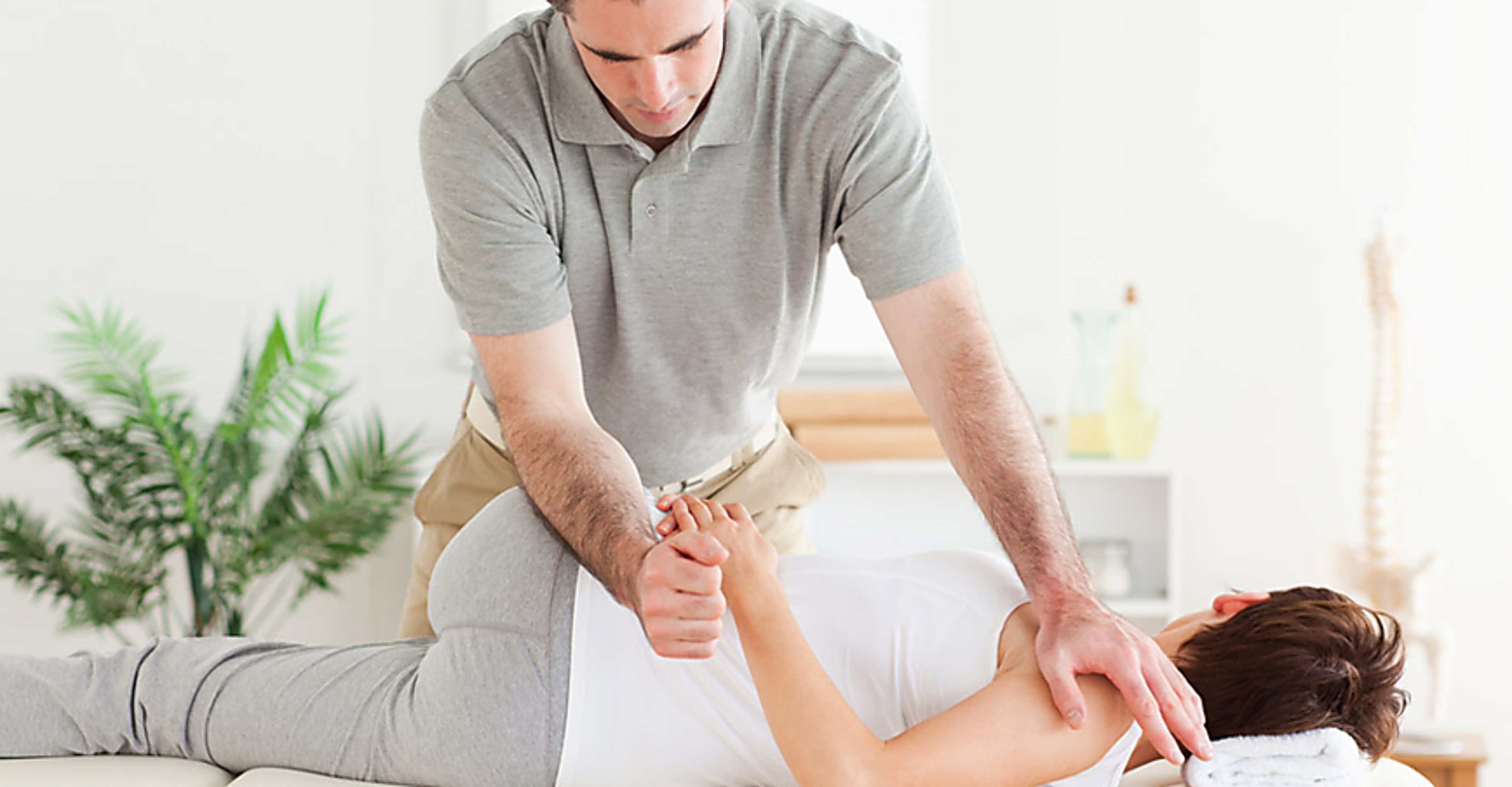
Lateral Elbow Pain
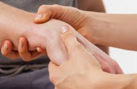
Lateral elbow pain is an extremely common presentation among sportspeople and manual workers. The most common cause is an overuse syndrome related to excessive wrist extension. This condition has traditionally been known as ‘tennis elbow’. This is an unsatisfactory term as it gives little indication of the pathological processes involved. In fact, the condition is more common in non-tennis players than in tennis players. It has also been referred to as ‘lateral epicondylitis’. This is also inappropriate as the site of the abnormality is usually just below the lateral epicondyle and the primary pathology is due to collagen disarray rather than inflammation.
The primary pathological process involved in this condition is tendinosis of the extensor carpiradials brevis (ECRB) tendon, usually within 1-2 cm of its attachment to the common extensor origin at the lateral epicondyle. This condition will be referred to as extensor tendinopathy.
Other conditions that may cause lateral elbow pain include synovitis of the radio humeral joint, radiohumeral bursitis and entrapment of the posterior interosseous branch of the radial nerve (radial tunnel syndrome). These conditions may exist by themselves or in conjunction with extensor tendinopathy.
There is often a contribution to lateral elbow pain from the cervical and upper thoracic spines and neural structures. This may be a relatively minor contribution or, in some cases, the main cause of the patients elbow pain. A full assessment of the cervical spine and neural structures is essential in examination of the patient with lateral elbow pain.
History
The characteristics of the patients lateral elbow pain should be elicited. The diffuse pain of extensor tendinopathy typically radiates from the lateral epicondyle into the proximal forearm extensor muscle mass. Occasionally the pain may be more localized. The onset of pain may be either acute or insidious. There may have been recent changes in training or technique, note-taking or equipment used in sport or work.
The severity of pain ranges from relatively trivial pain to an almost incapacitating pain that may keep the patient awake at night. It is important to note whether the pain is aggravated by relatively minor everyday activities, such as picking up a cup, or whether it requires repeated activity, such as playing tennis or bricklaying, to become painful.
Pain may radiate into the lateral aspect of the forearm. This may be consistent with posterior interosseous nerve entrapment or irritation of other neural structures. If pain is closely related to the activity level, it is more likely to be of a mechanical origin. If pain is persistent, unpredictable or related to posture, referred pain should be considered.
Certain movements, usually those involving wrist extension or gripping, will aggravate mechanical pain. Referred pain is affected by prolonged posture, such as lengthy periods seated at a desk or in a car. Associated sensory symptoms, such as pins and needles, may indicate a neural component. Presence of neck, upper thoracic or shoulder pain should also be noted.
Often by the time the patient presents to the sports medicine clinician, he or she will already have undergone a variety of treatments. It is important to note the response to each of these treatments.
An activity history should also be taken, noting any recent change in the level of activity. In tennis players, note any change in racquet size, grip size or string tension and whether or not any comment has been made regarding his or her technique.
Extensor tendinopathy
For this major sports medicine condition, we review the pathology, outline the clinical presentation, and then discuss evidence based and clinically founded treatment.
Clinical Features
Extensor tendinopathy occurs in association with any activity involving repeated wrist extension against resistance. This includes sporting activities, such as tennis ,squash and badminton, as well as occupational and leisure activities, such as carpentry, bricklaying, sewing and knitting. Computer use has been shown to be associated with the development of this condition. The peak incidence is between the ages of 40 and 50 years but this condition may affect any age group.
There are two distinct clinical presentations of this condition. The most common is an insidious onset of pain, which occurs 24-72 hours after unaccustomed activity involving repeated wrist extension. This occurs typically after a person spends the weekend laying bricks or using a screwdriver. It is also seen after prolonged sewing or knitting .In the tennis player, it may occur after the use of a new racquet, playing with wet, heavy balls or over hitting, especially hitting into the wind. It also occurs when the player is hitting ‘late’(getting the position slowly), so that body weight is not transferred correctly and the player relies on the forearm muscles exclusively for power.
Treatment
No single treatment has proven to be totally effective in the treatment of this condition. A combination of the different treatments mentioned below will result in resolution of the symptoms in nearly all cases.
The basic principles of treatment of soft tissues injuries apply. There must be control of pain, encouragement of the healing process, restoration of flexibility and strength, treatment of associated factors (e.g. increased neural tension, referred pain),gradual return to activity with added support and correction of the predisposing factors.
Control of Pain
It remains unclear as to how much pain is ideal in the treatment of tendinopathies. Clinical experience suggests that a low level of pain, which does not worsen with training, is likely to not be harmful for tendon healing. However, some patients require relative rest, application of ice and analgesia for comfort.


 Biceps Tendinitis