Tag Archives: Rehabilitation Services

How to Remove the disability of Bandy Legs?

Types, Causes and Treatment of Bandy Legs:

images 1Genu Varum  is defined as a lateral angulation of the knee. The longitudinal axis of femur and tibia deviates medially. The deformity involves tibia alone or the femur or tibia  and fibula both.

Types and Causes

Unilateral

  • Due to growth abnormalities of upper tibial epiphysis.
  • Infections like osteomyelitis, etc.
  • Trauma near the growth epiphysis of femur.
  • Tumors affecting the lower end of femur and upper end of tibia.

Bilateral

  • Congenital causes
  • Postural abnormalities
  • Developmental disorders
  • Metabolic disorders (rickets rare)
  • Endocrine disorders
  • Degenerative disorders (e.g.  Osteoarthritis of knee). This is a common cause.
  • Occupational disorders (e.g. in jockeys)
  • Idiopathic
  • Paget’s disease
  • ‘Blounts’ disease (tibia vara)

Clinical Measurements of the Deformity

Child

  • The patient is examined supine with knee extended, patella facing the ceiling and the medial malleoli touching each other. If the separation of the knee exceeds more than 3 cm or if it is unilateral, it should be investigated.
  • A line is drawn from anterosuperior iliac spine through center of patella to medial malleolus. Normally all the structures are in the same line but in genu varum medial malleolus is medial to this line.

Adults

The angle of genu varum is calculated on a standing radiograph of the whole limb.

Clinical Feature

Gena Varum complex: The primary deformity in genu varum is lateral angulation of the knee. In response to this, secondary deformities develop in the tibia and the foot. This together is known as genu varum complex.

Radiograph

Radiograph of the whole limb should be done to assess the severity of genu varum.

Treatment

  • Treatment should be conservative until four years of age. Knee-ankle-foot outhouses with the medial bar and the lateral strap are used.
  • Correction of early deformity is done by dynamic bracing or splints.
  • After four years, significant deformity should be corrected by surgery.
  • Lateral epiphyseal stapling when the child is within the growth period and supracondylar medial open or lateral closed wedge osteotomy is done after skeletal maturity.

Genu varum is a relatively common in kids. Idiopathic shin vara is the most typical of the pathologic circumstances that are associated with bowed legs; therapy techniques differ with the individual’s age and the level of illness and problems. Active Physical Therapy’s experienced dedicated physical therapists and talented clinical team then design individualized treatment plans to achieve the specific goals for each patient per your doctor’s expectation. Call now for Quick Appointment: 301-916-8540

http://www.active-physicaltherapy.com

What are the Causes of Ankle Joint Injury?

Causes, Clinical Features and Treatment of Ankle Injury:

imagesThe tibiofibular syndesmosis, consisting of the anterior and posterior inferior tibiofibular ligaments and interosseous membrane, maintains the joint between the distal tibia and fibula. It plays a dynamic role in ankle function.

Causes

  • Diastasis (separation) occurs with partial or complete rupture of the syndesmosis ligament.
  • Ruptures of the syndesmosis are rarely isolated injuries but generally occur in association with deltoid ligament injuries or, more frequently, with fractures of either the fibula or the posterior and medial malleoli.

Clinical Features

The classic presentation includes:

  • Anterior ankle pain following a moderate-to-severe ankle injury.
  • Tenderness on examination located at the anterior aspect of the syndesmosis and interosseus membrane.
  • Painful active external rotation of the foot. If there is severe disruption of the syndesmosis, the squeeze test is positive.

Investigations

  • Plain X-rays are recommended to exclude fractures and osseous avulsions.
  • Mortise views may reveal widening of the syndesmosis.
  • Stress X-rays in external rotation may demonstrate the diastasis.
  • CT or MRI is required to exclude osteochondral lesions.
  • Isotope bone scan may reveal a focal increased uptake in the region of the anterior tibiofibular ligament and interosseous membrane.

Treatment

  • Provided there is no widening of the distal tibiofibular joint, conservative management with rest, NSAIDs and physiotherapy is required.
  • As the pain settles, strengthening, range of motion and proprioceptive exercises are introduced.
  • In more severe cases, when there is widening of the distal tibiofibular joint, surgery and insertion of a temporary syndesmosis screw is required.

The shin and fibula are the two lengthy bone fragments of the reduced leg. Distal tibiofibular joint accidents typically happen traumatically during more serious rear-foot accidents whereby causes force the shin and fibular apart. Active Physical Therapy is recognized as a provider of superior care for orthopedic, Auto Accident, Sports Injuries / Trauma Cases, Work-related Injuries and comprehensive physical therapy services that improve function, encourage independence and better your quality of life. Call now for quick Appointment: 301-877-2323

Visit our Website: www.active-physicaltherapy.com

How to get comfort from Collateral Ligament Injury?

Clinical Features and Treatment of Collateral Ligament Injury (Knee Ligament):  

Collateral Ligament InjuryCollateral ligament injury is due to direct or indirect violence as described earlier. Medial collateral ligament injury is more common due to the valgus stress caused by striking the lateral aspect of the knee joint during collision in sports. The varus force on the medial side required to cause the lateral collateral ligament injury is less common because of the protection offered by the other leg.

Clinical Features

  • The patient gives history of valgus and external rotation force in mild sprains.
  • In severe sprains, the patient gives history of valgus stress force due to the direct blow on the lower thigh or upper leg seen commonly in contact sports like football, rugby, etc.
  • It may be associated with ACL tear or meniscal injury and then the patient may present with pain, swelling, hemarthrosis, etc.
  • On examination, the point of local tenderness could be at adductor tubercle, joint line or at the insertion of tibial collateral ligament.
  • About 10-20 percent of patients have damage to the extensor mechanism of the knee.

Investigations

  • Stress radiographs at 15-20 degree of valgus.
  • MRI helps to localize the MCL tears, ACL, meniscal injuries, etc.
  • Arthrograms and arthroscopy to evaluate and rule out meniscal and cruciate pathology.

Treatment

Fresh injury nonoperative treatment is the mainstay of treatment.

  • Sprain symptomatic treatment, nonsteroidal anti-inflammatory drugs (NSAIDs), etc.
  • Sprain long leg cast for 4-6 weeks with knee in 30-40 of flexion.
  • Sprain surgical repair in isolated tears. Repair and reconstruction in old tears or in associated injuries. Brace is required for 4-7 months.

The knee is the biggest joint in your body and one of the most complicated. It is also vital to activity. Your knee structures link your thighbone to your lower leg bone fragments. Knee structures injuries or holes are a common activities injury. Athletes who get involved in immediate contact activities like soccer or soccer are more likely to harm their security structures. Active Physical Therapy is recognized as a provider of superior care for orthopedic, Auto Accident, Sports Injuries / Trauma Cases, Work-related Injuries and comprehensive physical therapy services that improve function, encourage independence and better your quality of life. Call now for best Physical Therapy: 301-662-9335

http://www.active-physicaltherapy.com

How should we prevent coronoid fracture?

Classification And Treatment of Elbow Fractures:Coronoid FractureFractures of the coronoid process of the ulna were earlier thought to be an avulsion fracture involving the brachial is muscle. Of late, this notion has been dispelled as it is found that the insertion of this muscle is more distal.

Interesting Facts About Coronoid Fractures

  • Its presence indicates a significant trauma to the elbow.
  • It also points towards the possibility of acute recurrent dislocations.

Mechanism of Injury

This fracture occurs due to the impact of the coronoid process against the trochlea following a fall on an outstretched hand.

Classification of Regan and Morrey

  • Type I: Avulsion fracture of the tip of the coronoid.
  • Type II: Fracture involving greater than 50 percent of the coronoid.
  • Type III: Fracture involving the base of the coronoid.

Clinical Features

Isolated fractures of the coronoid process are usually rare and are usually associated with greater elbow trauma. Clinical features like pain, swelling, deformity, movement restriction of the elbow, etc. depends on the extent of damage.

Radiograph

This fracture can be easily identified over a true lateral X-ray of the elbow.

Treatment

  • Though small-undisplaced fractures can be managed conservatively with an above elbow plaster cast, displaced fractures need open reduction and internal fixation with screw or wires.
  • A physician might be able to reattach a division or repair a damaged bone, but it often takes actual recovery to restore a person’s function.
  • Physical Therapy treatment, in most cases, contains direct modification of muscles, joint parts and other parts of the body affected by a harm or serious sickness.
  • It often contains body building, heat treatments, massages therapy and supervised exercises.
  • Individual workouts often depend on the type of harm or condition, the person’s age and specific treatments recommended by a doctor.

If you are suffering from elbow fracture then come instantly our center Active Physical Therapy. For more detailed information Call Now at: 301-498-1604.

http://www.active-physicaltherapy.com

How to Cure Toe Arthritis?

Causes and Treatment of Hallux Limitus (Toe Arthritis):

Hallux Limitus is defined as a restriction in dorsiflexion of the hallux at the first metatarsophalangeal joint secondary to exostoses or osteoarthritis of the joint. Often the term ‘hallux rigidus’ is used to describe the final progression of hallux limitus as ankylosis of the joint occurs.

The primary role of the hallux is to enable dorsiflexion of the first metatarsal during the propulsive phase of gait. Limitation of this range of motion results in problems with gait.

Causes

  • Trauma-secondary to chondral damage
  • Excessive pronation of the foot may increase the stresses on the joint and promote development of exostoses
  • Repetitive weight-bearing dorsiflexion of the first metatarsophalangeal joint
  • Autoimmune arthropathy (e.g. rheumatoid arthritis)
  • Aberration of the first metatarsal or proximal phalanx
  • Hypermobile first ray
  • Muscle imbalance

Hallux LimitusClinical Features

  • The main presenting symptom is usually that of pain around the first metatarsophalangeal joint. The pain is often described as a deep aching sensation that is aggravated by walking, especially in high heels, or activities involving forefoot weight-bearing.
  • Dorsal joint hypertrophy can be a source of irritation from footwear and may lead to pain secondary to skin or soft tissue irritation.
  • In patients with longstanding hallux limitus, a distinct shoe wear pattern is seen: the sole demonstrates wear beneath the second metatarsophalangeal joint and the first interphalangeal joint.
  • Examination reveals tenderness of the first metatarsophalanageal joint, especially over the dorsal aspect, often with palpable dorsal exostoses.
  • There is a painful limitation of joint motion, the degree of limitation reflecting the severity of the arthrosis.

Investigations

  • Plain X-rays display the classic characteristics of degenerative osteoarthritis and the degree of degeneration observed will reflect the duration and severity of the condition.
  • Features include joint space narrowing, sclerosis of the subchondral bone plate, osteophytic proliferation, flattening of the joint, sesamoid displacement and free bony fragments.

Treatment

  • Conservative management consists of an initial reduction in activity, NSAIDs, a cortisone injection if required, physiotherapy, and correction of biomechanical factors with orthoses and/or footwear.
  • Conservative treatment often fails when hallux dorsiflexion is less than 50 degree. In extreme cases, cheilectomy is required.
  • Occasionally, arthroplasty of the first metatarsophalangeal joint is indicated.

Hallux Limitus is basically restricted movement of the big toe combined. The big toe combined generally will go through a 55-65 level variety of flexibility. With Hallux Limitus this movement may be decreased to 25 or 30 levels. Hallux Rigidus is a firm movement or finish lack of any movement of this combined. As like a bunion problems this is a cuboid architectural problems. If you are suffering from Toe Arthritis then call at Active Physical Therapy301-916-8540

Visit Our Website: http://www.active-physicaltherapy.com

Treatment of Intra-Articular Fractures of The Calcaneus

Clinical Features And Treatment of Heel Bone Fracture:INTRA ARTICULAR FRACTURESThese account for 60 percent of all tarsal injuries and 75 percent of all calcaneal fractures.

Mechanism of Injury

Fall  From  Height:  Lateral  process  of  talus  acts  as  a wedge  and  is forced  through  the  Gissane’s  angle resulting in four fracture  patterns:

  • Undisplaced
  • Tongue shaped
  • Joint  depression
  • Comminuted

Clinical Features

Pain and swelling of the heel, the patient is unable to bear weight, stand or walk, pain and difficulty during inversion and eversion of the heel.

Clinical Signs

  • Swelling over the heel.
  • Tenderness over the heel.
  • Lateral  heel  compression  test  elicits  pain
  • Broadening of the heel
  • Horseshoe swelling on either side the  tendo-Achilles
  • Distance between the heel and malleoli is reduced.

Radiography

  • Plain X-rays of the foot as in extra-articular fractures.
  • CT scan is now emerging as the gold standard in evaluation of intra-articular calcaneal fractures.

Treatment

1)      CONSERVATIVE

The following are the basic methods of treatment:

  • No reduction and early motion consists
  • Elastocrepe bandage application
  • Foot elevation
  • Weight bearing at the end of 12 weeks
  • Closed reduction and fixation.

2)    GOALS

Common Steps of Reduction:

  • Under anesthesia (general or spinal), the patient is prone and knee is flexed to 90 degree.
  • With the assistant supporting the thigh, the surgeon compresses the medial and lateral sides of the heel.
  • Strong longitudinal traction is now applied along the direction of the leg.
  • Varus or valgus force is now applied depending on the displacement.
  • Lastly the calceneal tuberosity is manipulated in position.
  • Compression bandage is finally applied.

Surgery

Severely comminuted and depressed fracture with subchondral defects requires open reduction and internal fixation with cancellous bone graft to fill the gap. Recently, for this purpose, alternatively, biocompatible and less re-absorbable nanocrystalline calcium phosphate cement called Bioban is being tried with successful results in some centers. Open reduction and internal fixation with plate and screws are difficult and are rarely adopted.

Complications

  • Nonunion is rare due to the cancellous nature of the bone.
  • Malunion is more common.
  • Heel Pain: The source of heel pain could be from:
  1. Subtalar joint due to post-traumatic osteoarthritis.
  2. Peroneal tendonitis due to stenosing.
  3. Tenovaginitis of the peroneal tendons.
  4. Bone spurs due to Malunion of fracture.
  5. Disruption of fat pad of the heel.
  6. Arthritis of calcaneocuboid joint is a major source of pain.
  7. Nerve entrapment is rare. Medial or lateral plantar branches of posterior tibial nerve or sural nerve may be entrapped due to soft tissue scarring.

If you are being effected from this fracture come instantly our center Active Physical Therapy. For more information or questions, feel free to contact any of our offices located in Maryland. You can also make your appointment online to start your treatment within 24 to 48 hours at Active Physical Therapy. Call Now at: 301-498-1604

http://www.active-physicaltherapy.com/

Ilipsoas-Related Longstanding Groin Pain

Clinical Features And Treatment of Groin Pain:ILIPSOAS-RELATED LONGSTANDING GROIN PAINThe Iliopsoas muscles may be the sole cause of the athletes longstanding groin pain, but this component is frequently present in conjunction with adductor abnormalities. The Iliopsoas component needs to be recognized and subsequently treated.

The Iliopsoas muscle is the strongest flexor of the hip joint. It arises from the five lumbar vertebrae and the ilium and inserts into the lesser trochanter of the femur. It is occasionally injured acutely but frequently becomes tight with neural restriction. Whether or not Iliopsoas tendinopathy and bursitis contribute substantially to exercise-related groin pain remains unclear. Most case reports associate these conditions with hip surgery and with rheumatological conditions (e.g. polymyalgia rheumatic). The thin-walled Iliopsoas bursa commonly communicates with the hip joint. Experienced clinicians feel that muscular and neuromyo fascial elements are likely to contribute far more commonly than do Iliopsoas bursitis and tendinopathy.

Clinical Features

Iliopsoas problems may occur as an overuse injury resulting from excessive hip flexion, such as kicking. They present as a poorly localized ache that patients usually describe as being a deep ache in one side of the groin. There are two key clinical signs that point to the Iliopsoas as the source of groin pain.

  • The first, tenderness of the muscle in the lower abdomen, relies on palpation of the Iliopsoas muscle, which is difficult in its proximal portion, deep within the pelvis.
  • Nevertheless, the skilled examiner may detect tenderness more distally, particularly in thin athletes, by palpating carefully just below the inguinal ligament, lateral to the femoral artery and medial to the Sartorius muscles.
  • Passive hip flexion facilitates this palpation.
  • The second key clinical sign that helps distinguish the Iliopsoas from other sources of groin pain is pain and tightness on Iliopsoas stretch that is exacerbated on resisted hip flexion in the stretch position.
  • Frequently, the further addition of passive cervical flexion and knee flexion will aggravate the pain, indicating a degree of neural restriction through the muscle.
  • It is important to examine the lumbar spine as there is frequently an association between Iliopsoas tightness and hypomobility of the upper lumbar spine from which the muscle originates.

Treatment

Treatment of Iliopsoas-related groin pain is similar to that of adductor-related groin pain but with an increased emphasis on soft tissue treatment of the Iliopsoas and Iliopsoas stretching with the addition of a neural component. Often, mobilization of the lumbar intervertebral joints at the origin of the Iliopsoas muscles will markedly decrease the patient’s pain.

The article presents clinical examination of athletes with groin pain. Clinical examination techniques that are used to diagnose and evaluate the degree of groin pain in athletes have not been well evaluated.Active Physical Therapy provides our services include and are not limited to physical therapy, occupational therapy, hand therapy, senior wellness, neurological rehabilitation, orthopedic rehabilitation, industrial rehabilitation and specialties including auto accident injuries / Trauma Cases, Work-related Injuries, Sports Injuries, Tennis Elbow,etc. For More Detailed Information Call Now at: 301-498-1604

http://www.active-physicaltherapy.com/

How to Cure Harmed Tendons?

Examination and Treatment of Achilles Tendon:       Achilles Tendon

Acute tendon rupture is most common among men aged 30-50 years (mean age, 40 years) it causes sudden severe disability.

Achilles tendon injuries tendinopathy may arise with increased training volume or intensity but may also arise insidiously. Because the prognosis for midportion Achilles tendinopathy is much better than for insertional tendinopathy, these conditions should be distinguished clinically. The condition that was previously called ‘Achilles tendinitis‘ is not truly an inflammatory condition and, thus, should be referred to as ‘Achilles tendinopathy’. The main differential diagnoses of gradual onset pain in the Achilles region arise from the neighboring anatomy.

There are two bursae in this region:

  • The Retrocalcaneal Bursa
  • The Achilles Bursa

1.   The Retrocalcaneal Bursa: Retrocalcaneal Bursa lies between the posterior aspect of the calcaneus and the insertion of the Achilles tendon.

2.   The Achilles Bursa: Achilles Bursa lies between the insersion of the Achilles tendon and the skin.

The posterior process of the talus or a discrete anatomical variant, the os trigonum, can each be involved in posterior impingement syndrome. This is most commonly seen in ballet dancers but occurs occasionally in sprinters and in football players. Other, much less common differential diagnoses include dislocation of the peroneal tendons, an accessory soleus muscle, irritation, or neuroma of the sural nerve, and systemic inflammatory disease. These pathologies cause pain in and also around the Achilles tendon; true tendon pain is almost always confined to the tendon itself. In adolescents, it is important to consider the diagnosis of Sever’s lesion, a traction apophysitis at the insertion of the Achilles tendon into the calcaneus. Referred pain is a very rare cause of pain in the Achilles region.

History

  • The athlete with overuse tendinopathy notices a gradual development of symptoms and typically complains of pain and morning stiffness after increasing activity level.
  • Pain diminishes with walking about or applying heat. In most cases, pain diminishes during training, only to recur several hours afterwards.
  • The onset of pain is usually more sudden in a partial tear of the Achilles tendon. In this uncommon condition, pain may be more disabling in the short term.
  • As the histological abnormality in a partial tear and in overuse tendinopathy are identical, we do not emphasize the distinction other than to suggest that time to recovery may be longer in cases of partial tear.

Examination

Palpate the painful area for tenderness, thickening, and crepitus. If the Achilles tendon seems to be the cause of pain, and the examiner is confident that the tendon is intact, the examination should aim to provoke tendon pain during tendon loading activity.  These functional tests provide a baseline against which treatment response can be compared. Another method of monitoring the clinical progress of Achilles tendinopathy is to use the VISA questionnaire. This is simple questionnaire takes less than 5 minutes to complete and once patients are familiar with it they can complete most of it themselves.

Examination involves:

1.   Observation

  • Standing
  • Walking
  • Prone

2.   Active movements

3.   Passive movements

  • Plantarflexion
  • Plantarflexion with overpressure
  • Dorsiflexion
  • Subtalar joint
  • Muscle stretch
  • Gastrocnemius
  • Soleus

4.   Resisted movements

  • Plantarflexion- calf raises

5.   Functional tests

  • Single- leg calf raises
  • Hop
  • Eccentric drop

6.   Palpation

  • Achilles tendon
  • Retrocalcaneal bursa
  • Posterior talus
  • Calf muscle

7.   Special test

  • Prone inspection for tendon rupture
  • Simmond’s Calf squeeze test
  • Biomechanical assessment

Investigations and Treatment of Achilles Tendons

  • Plain radiographs are of limited value but, if symptoms are longstanding, they may reveal a Haglund’s deformity, a prominent superior projection of the calcaneus, or spurs projecting into the tendon.
  • Posterior impingement can be shown radiographically using functional views. X-ray may reveal calcification in the tendon itself but, unless severe, this can be asymptomatic.
  • In symptomatic patients, both ultrasound and MRI often reveal an abnormal signal in the Achilles tendon that generally corresponds with the histopathology of tendinosis.
  • Ultrasound and MRI can help distinguish different causes of pain in the Achilles region.
  • Achilles tendon insertion is abnormal in patients with pain at the distal tendon.
  • It may also provide a target for treatment. Because of the variability in imaging and its inconsistent clinical correlation, the results of imaging should not dominate clinical decision making variation in symptoms such as morning stiffness and load pain should direct treatment modification. Studies in many tendons have indicated that clinical outcomes are independent of imaging and change in imaging.

Achilles tendinitis is a typical situation that causes discomfort along the returning of the leg near the returning heel. Active Physical Therapy is the best Physical Therapy Center which is specialists in treating Ankle Sprains,  Knee and Ankle Injuries, Bursitis/Tendonitis etc. Call now at: 301-662-9335

http://www.active-physicaltherapy.com

Patellofemoral Pain (Runner’s Knee)

Treatment of Knee Pain:

Definition of Patellofemoral PainPatellofemoral Pain

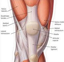
Patellofemoral pain is the preferred term used to describe pain in and around the patella. Synonyms include PFJ syndrome, anterior knee pain and chondromalacia patellae.

Patellofemoral pain is an umbrella term used to embrace all peripatellar or retro patellar pain in the absence of other pathologies. Since the cause of the pain may differ between patients, it is appropriate to review the potential sources of patellofemoral pain. A number of extra- and intra-articular components of the knee can generate neurosensory signals that ultimately result in of the patient feeling pain. Patellofemoral articular cartilage cannot directly be a source of pain.
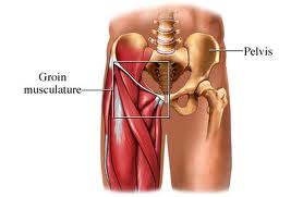
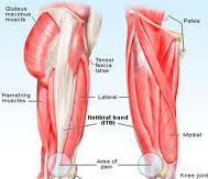
Functional Anatomy

  • At full extension, the patella sits lateral to the trochlea.
  • During flexion, the patella moves medially and comes to lie within the Intercondylar notch until 130 degree of flexion, when it starts to move laterally again.
  • The patell mediolateral excursion is controlled by the quadriceps muscles, particularly the VMO and vastus lateralis components.
  • With increasing knee flexion, a greater area of patellar articular surface comes into contact with the femur, thus offsetting the increased load that occurs with flexion.
  • Loaded knee flexion activities subject the PFJ to loads many times the body weight, ranging from 0.5 times body weight for level walking to seven to eight time’s body weight for stair climbing.

Factors That May Contribute To Development of Pain

  1. Remote Contributing Factors
  2. Local Contributing Factors

Increased PFJ load instigates the development of patellofemoral pain. Factors that influence PFJ load can be considered in two categories: extrinsic and intrinsic.

  • During physical activities the extrinsic load is created by the body’s contact with the ground (ground reaction force) and is therefore moderated by body mass, speed of gait, surfaces and footwear.
  • During weight-bearing activities, any increase in the amount of knee flexion will increase the PFJ load.
  • Intrinsic factors can influence both the magnitude and the distribution of the PFJ load.
  • Distribution of load is conceptualized as movement of the patella within the femoral trochlea: patella tracking.
  • Local factors that influence patella movement include patella position, soft tissue tension and neuromuscular control of the medial and lateral components of the vasti.
  • The clinician should assess the contribution of various extrinsic and intrinsic factors to the development of patellofemoral pain.
  • This assessment is crucial in the planning of an appropriate treatment regimen.
  • The history will elucidate valuable information pertaining to extrinsic factors but clinical examination is usually required to evaluate most intrinsic contributing remote and local factors.

1. Remote Contributing Factors

The following remote factors may contribute to the development of patellofemoral pain:

  • Increased femoral internal rotation
  • Increased knee valgus
  • Increased tibial rotation
  • Increased subtalar pronation
  • Inadequate flexibility

It is important to assess the patient in static postures as well as functional activities. Some factors may become more obvious during specific functional tasks, such as the step-down or single-leg squat, where the postural demands are high.

2. Local Contributing Factors

Local factors that can contribute to the development of patellofemoral pain are:

  • Patella position
  • Soft tissue contributions
  • Neuromuscular control of the vasti

Treatment of Patellofemoral Pain

The management of a patient with patellofemoral pain requires an integrated approach that may include:

  • Reduction of pain and inflammation
  • Addressing extrinsic contributing factors
  • Addressing intrinsic contributing factors:
  • Evidence base for physical intervenons
  • Surgery-to be avoided

Immediate Reduction of Pain

The first priority of treatment is to reduce pain. This may require some or all of the following: rest from aggravating activities, ice, a short course of NSAIDs, electrotherapeutic modalities (e.g. ultrasound) and techniques such as mobilization or dry needling or acupuncture. Taping should have an immediate pain-relieving effect.

Addressing Extrinsic Contributing Factors

While initially it is vital to advise the patient to reduce the load on the PFJ, as rehabilitation progresses it is essential that any extrinsic factors that may have been placing excessive load on the PFJ (e.g. training, shoes and surfaces) are discussed and modified if necessary.

Addressing Intrinsic Contributing Factors

The clinician should have ascertained from the outset whether any intrinsic factors may have contributed to the development of the patient’s pain. Remote intrinsic factors may be addressed through hip muscle retraining, improving musculotendinous compliance or foot orthoses. Local intrinsic factors may be addressed with techniques such as patella taping or bracing, improving lateral soft tissue compliance, generalized quadriceps strengthening or vasti retraining.

Evidence Base for Physical Intervenons

A number of controlled clinical trials have assessed the effectiveness or efficacy of physical interventions for patellofemoral pain. While these reflect some aspects of treatment techniques, mostly the interventions are not individualized to the patients needs.

Treatment options that have gained popularity more recently have not been evaluated as thoroughly. The level 1 (systematic reviews) and level 2 (controlled clinical trials) evidence of the evaluation of physical interventions for patellofemoral pain.

Surgery-To Be Avoided

Experienced clinicians will have observed that the need for surgery in patellofemoral pain has been greatly reduced. This is likely due to the availability of evidence-based, exercise-based, physical interventions. To our knowledge, there has been no surgical randomized controlled trial showing the effectiveness of treatments such as chondroplasty or lateral release for patellofemoral pain. Thus, at a time when systematic reviews (level evidence) argue for physical therapies for this condition, it would appear that such avenues should be tried repeatedly and with various expert physical therapists before being abandoned in favor of a hoped-for surgical miracle. We note that poor surgical outcomes have been reported and often patellofemoral pain is worsened after surgery.

Active Physical Therapy’s experienced dedicated physical therapists and talented clinical team then design individualized treatment plans to achieve the specific goals for each patient per your doctor’s expectation.For More Information Call Now at: 301-498-1604

http://www.active-physicaltherapy.com/

Are you being affected by low back pain?

Severe Low Back Pain Treatment

The majority of patients with low back pain present with mild-to-moderate pain. A small group of patients present with acute onset of severe low back pain.

The aim of initial management of these patients is to reduce the pain and inflammation as rapidly as possible. When this is done, the management of these patients relies on the same principles as those with mild-to-moderate low back pain.

Acute onset of severe low back pain in the absence of nerve root signs may be due either to an acute tear of the anulus fibrosus of the disk or to an acute locked apophyseal joint. A locked apophyseal joint is thought to be due to entrapment of an intra-articular meniscus.

Clinical Features of Severe Acute Low Back Pain

  • Acute low back pain is usually of sudden onset and is often triggered by a relatively minor movement such as bending to pick up an object.
  • This minor incident may be more indicative of fatigue or lack of control, rather than tissue overload. The pain may increase over a period of hours due to the development of inflammation.
  • Patients with chronic low back pain may also have acute exacerbations that may become more frequent and require less initiation over time.
  • The pain is usually in the lower lumbar area and may be central, bilateral or unilateral.
  • It may radiate to the buttocks, hamstrings or lower leg. Sharp, laminating pain in a narrow band down the leg is radicular pain and is associated with nerve root irritation, commonly as a result of intervertebral disk prolapsed.
  • More commonly, the pain referred to the buttock and hamstring is somatic in nature, with the patient complaining of a deep-seated ache.
  • The patient with acute, sudden onset of low back pain often adopts a fixed position and movements are severely restricted in all directions.
  • Palpation of the lumbar spine reveals areas of marked tenderness with associated muscle spasm.

Management of Severe Acute Low Back Pain

  • Encourage the patient to adopt the position of most comfort position varies considerably and may be lying prone, supine or, commonly, side-lying with a degree of lumbar flexion.
  • Movements that aggravate pain should be avoided, whereas movements that reduce or have no effect on pain should be encouraged.
  • Bed rest in the position of most comfort may be continued for up to 48 hours depending on the amount of pain.
  • Bed rest longer than 48 hours has been shown to be detrimental.
  • Taping of the low back can markedly reduce acute back pain and allow quicker functional restoration.
  • Analgesics may control the pain and reflex muscle spasm. NSAIDs may help reduce inflammation.
  • Electrotherapeutic modalities, for example, TENS, interferential stimulation and magnetic field therapy, may be helpful in reducing pain and muscle spasm in the acute stage. However, if access to these modalities in the acute stage requires any degree of travel, then bed rest alone may be preferable.
  • Exercise in a direction away from the movement that aggravates the patient’s symptoms should be commenced as early as possible. For those patients in whom flexion aggravates their symptoms, extension exercises should be performed.
  • The degree of extension should be determined by the level of pain. Initially, lying prone may be sufficient. Later, extension of the lumbar spine by pushing up onto the elbows may be possible. Eventually, further extension with straight arms can be achieved.
  • Exercises should be immediately discontinued if peripheral symptoms develop.
  • Prolonged posture involving flexion, such as sitting, should be avoided.
  • In patients for whom extension movements aggravate their pain, flexion exercises or rotation (away from pain) exercises should be performed. For these patients, prolonged posture involving extension, such as standing with excessive lumbar lordosis, should be avoided.

Manual therapy has only a limited role in treating severe low back pain. Gentle mobilization techniques, for example, posteroanterior (PA) mobilization, may be performed and the patient’s response closely monitored. If there is any deterioration of symptoms, mobilization should be immediately ceased. The mobilization should be performed in the position of comfort adopted by the patient. Manipulation should not be attempted in the presence of marked muscle spasm. Similarly, gentle (grade I) soft tissue massage may be helpful in relieving pain and muscle spasm Traction has not been found to be helpful in patients with acute low back pain.

If you are being affected by Severe Low Back Pain and want to get rid of this pain then search actual rehabilitation medical center near you and seek advice from only professional, certified and experienced physical therapist today. For More Information Call Now At:  301-498-1604

http://www.active-physicaltherapy.com/