Monthly Archives: March 2015

What are the causes of Knee Injury?

Causes and Treatment of Knee Injury:

Osteochondritis dissecans is a combined condition whereby a varying amount of cuboid and its nearby fibrous drops its blood flow. Osteochondritis dissecans can include the cuboid and fibrous of almost any combined. Joints are most commonly affected. In this condition, avascular necrosis occurs in an area of subchondral bone followed usually by degenerative changes in the overlying cartilage. Though it can occur in any joint, it is most commonly seen in the knee joint. This avascular bone undergoes necrosis, gets detached and forms a loose body. Infact, this is the most common cause for loose bodies within the knee.

Knee InjuryCauses

Many causes are cited and are controversial:

  • Exogenous trauma
  • Endogenous trauma
  • Ischemia
  • Abnormal ossification within the epiphysis
  • Genetics
  • Combination of these
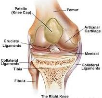
Common site

Lateral aspect of the medial femoral condyle near the attachment of posterior cruciate ligament.

Age groups

  • In young patients before epiphyseal closure. Treatment outcome is good.
  • In adults, here treatment outcome is poor.

Clinical Features

  • It is different in the two age groups and consists of vague pain and discomfort in the knee, swelling, catching, popping and locking could occur.
  • After complete separation, loose bodies can be palpated.
  • Tenderness can be elicited over the anteromedial surface of the femoral condyle by deep palpation after flexing the knee.

Investigations

Plain X-rays of the knee (AP, lateral and tunnel view), arthroscopy, bone scan, MRI, etc. are some of the important investigation tools. Plain X-ray also helps to detect the loose bodies of the knee joints.

Treatment

  • This depends on the age of the patient and degree of involvement.
  • Treatment method varies from conservative in children to operative in adults.
  • The operative methods are arthroscopic excision, curettage, pinning, debridement, grafting, etc.
  • The outcome of the treatment is good in children and is not so good in adults.

If you are suffering from Knee Injury then contact today for Quick Appointment at Active Physical Therapy. Active Physical Therapy is the best Physical Therapy Clinic in Maryland.

http://www.active-physicaltherapy.com/

What treatment should be done for Hand Joint Pain?

Treatment of Hand Joint Pain:

Volar BartonVolar Barton (Palmar rim dislocation) is a palmar rim fracture of distal radius. The most common cause of this type of fracture is a drop on an outstretched hand.

Mechanism

It is due to palmar tensile stress and dorsal shear stress and is usually combined with Radial styloid fracture.

Clinical Features

Radiograph

Palmar rim of distal radial articular surface is displaced dorsally. Proximally and posteriorly and may be associated with dorsal subluxation of the wrist.

Treatment

Conservative

Reduction is simple, but retention is difficult. Long arm cast is used.

Surgery

If reduction does not remain satisfactorily with wrist in neutral or slight palmar flexion, fixation with K-wire, external fixators and buttress plate, etc. may be done. Ellis T-’shaped buttress plate fixation is the preferred method of treatment.

Active Physical Therapy provides state of the art physical therapy throughout the state of Maryland.  Active’s friendly staff looks forward to assisting you in making your appointment in any of our clinics. Contact Us at: 301-498-1604

http://www.active-physicaltherapy.com

How to prevent Hand Disorder?

Types and Treatment of Hand Injury:

indexThis is the fourth most common carpal bone fractures (after scaphoid, capitate and lunate).

Mechanism of Injury

Types of Hand Fracture

These are divided into two types:

  • Body fractures
  • Peripheral chip or avulsion fractures

Clinical Features

Investigations

Plain X-ray of the wrist (AP, lateral or oblique views) may reveal the fracture. If still in doubt, CT scan is recommended.

Treatment

Body Fractures

  • If undisplaced, it is treated by short arm cast for 4 to 6 weeks.
  • If displaced (> 1-2 mm), surgery is the treatment of choice namely:
  • Arthroscopic approach and percutaneus pinning.
  • Open reduction and internal fixation through K-wires or screws.

Chip or Avulsion Fractures

Symptomatic treatment by orthotics in most of the cases helps and rarely may be required in intractable cases.

If you are in Pain and Suffering from Hand Disorder then Visit now at Active Physical Therapy. Please do not hesitate to contact us, we are dedicated to providing the best physical therapy service in Maryland. Call at: 301-877-2323

http://www.active-physicaltherapy.com

How to Remove the disability of Bandy Legs?

Types, Causes and Treatment of Bandy Legs:

images 1Genu Varum  is defined as a lateral angulation of the knee. The longitudinal axis of femur and tibia deviates medially. The deformity involves tibia alone or the femur or tibia  and fibula both.

Types and Causes

Unilateral

  • Due to growth abnormalities of upper tibial epiphysis.
  • Infections like osteomyelitis, etc.
  • Trauma near the growth epiphysis of femur.
  • Tumors affecting the lower end of femur and upper end of tibia.

Bilateral

  • Congenital causes
  • Postural abnormalities
  • Developmental disorders
  • Metabolic disorders (rickets rare)
  • Endocrine disorders
  • Degenerative disorders (e.g.  Osteoarthritis of knee). This is a common cause.
  • Occupational disorders (e.g. in jockeys)
  • Idiopathic
  • Paget’s disease
  • ‘Blounts’ disease (tibia vara)

Clinical Measurements of the Deformity

Child

  • The patient is examined supine with knee extended, patella facing the ceiling and the medial malleoli touching each other. If the separation of the knee exceeds more than 3 cm or if it is unilateral, it should be investigated.
  • A line is drawn from anterosuperior iliac spine through center of patella to medial malleolus. Normally all the structures are in the same line but in genu varum medial malleolus is medial to this line.

Adults

The angle of genu varum is calculated on a standing radiograph of the whole limb.

Clinical Feature

Gena Varum complex: The primary deformity in genu varum is lateral angulation of the knee. In response to this, secondary deformities develop in the tibia and the foot. This together is known as genu varum complex.

Radiograph

Radiograph of the whole limb should be done to assess the severity of genu varum.

Treatment

  • Treatment should be conservative until four years of age. Knee-ankle-foot outhouses with the medial bar and the lateral strap are used.
  • Correction of early deformity is done by dynamic bracing or splints.
  • After four years, significant deformity should be corrected by surgery.
  • Lateral epiphyseal stapling when the child is within the growth period and supracondylar medial open or lateral closed wedge osteotomy is done after skeletal maturity.

Genu varum is a relatively common in kids. Idiopathic shin vara is the most typical of the pathologic circumstances that are associated with bowed legs; therapy techniques differ with the individual’s age and the level of illness and problems. Active Physical Therapy’s experienced dedicated physical therapists and talented clinical team then design individualized treatment plans to achieve the specific goals for each patient per your doctor’s expectation. Call now for Quick Appointment: 301-916-8540

http://www.active-physicaltherapy.com

What are the Causes of Ankle Joint Injury?

Causes, Clinical Features and Treatment of Ankle Injury:

imagesThe tibiofibular syndesmosis, consisting of the anterior and posterior inferior tibiofibular ligaments and interosseous membrane, maintains the joint between the distal tibia and fibula. It plays a dynamic role in ankle function.

Causes

  • Diastasis (separation) occurs with partial or complete rupture of the syndesmosis ligament.
  • Ruptures of the syndesmosis are rarely isolated injuries but generally occur in association with deltoid ligament injuries or, more frequently, with fractures of either the fibula or the posterior and medial malleoli.

Clinical Features

The classic presentation includes:

  • Anterior ankle pain following a moderate-to-severe ankle injury.
  • Tenderness on examination located at the anterior aspect of the syndesmosis and interosseus membrane.
  • Painful active external rotation of the foot. If there is severe disruption of the syndesmosis, the squeeze test is positive.

Investigations

  • Plain X-rays are recommended to exclude fractures and osseous avulsions.
  • Mortise views may reveal widening of the syndesmosis.
  • Stress X-rays in external rotation may demonstrate the diastasis.
  • CT or MRI is required to exclude osteochondral lesions.
  • Isotope bone scan may reveal a focal increased uptake in the region of the anterior tibiofibular ligament and interosseous membrane.

Treatment

  • Provided there is no widening of the distal tibiofibular joint, conservative management with rest, NSAIDs and physiotherapy is required.
  • As the pain settles, strengthening, range of motion and proprioceptive exercises are introduced.
  • In more severe cases, when there is widening of the distal tibiofibular joint, surgery and insertion of a temporary syndesmosis screw is required.

The shin and fibula are the two lengthy bone fragments of the reduced leg. Distal tibiofibular joint accidents typically happen traumatically during more serious rear-foot accidents whereby causes force the shin and fibular apart. Active Physical Therapy is recognized as a provider of superior care for orthopedic, Auto Accident, Sports Injuries / Trauma Cases, Work-related Injuries and comprehensive physical therapy services that improve function, encourage independence and better your quality of life. Call now for quick Appointment: 301-877-2323

Visit our Website: www.active-physicaltherapy.com

How to get comfort from Collateral Ligament Injury?

Clinical Features and Treatment of Collateral Ligament Injury (Knee Ligament):  

Collateral Ligament InjuryCollateral ligament injury is due to direct or indirect violence as described earlier. Medial collateral ligament injury is more common due to the valgus stress caused by striking the lateral aspect of the knee joint during collision in sports. The varus force on the medial side required to cause the lateral collateral ligament injury is less common because of the protection offered by the other leg.

Clinical Features

  • The patient gives history of valgus and external rotation force in mild sprains.
  • In severe sprains, the patient gives history of valgus stress force due to the direct blow on the lower thigh or upper leg seen commonly in contact sports like football, rugby, etc.
  • It may be associated with ACL tear or meniscal injury and then the patient may present with pain, swelling, hemarthrosis, etc.
  • On examination, the point of local tenderness could be at adductor tubercle, joint line or at the insertion of tibial collateral ligament.
  • About 10-20 percent of patients have damage to the extensor mechanism of the knee.

Investigations

  • Stress radiographs at 15-20 degree of valgus.
  • MRI helps to localize the MCL tears, ACL, meniscal injuries, etc.
  • Arthrograms and arthroscopy to evaluate and rule out meniscal and cruciate pathology.

Treatment

Fresh injury nonoperative treatment is the mainstay of treatment.

  • Sprain symptomatic treatment, nonsteroidal anti-inflammatory drugs (NSAIDs), etc.
  • Sprain long leg cast for 4-6 weeks with knee in 30-40 of flexion.
  • Sprain surgical repair in isolated tears. Repair and reconstruction in old tears or in associated injuries. Brace is required for 4-7 months.

The knee is the biggest joint in your body and one of the most complicated. It is also vital to activity. Your knee structures link your thighbone to your lower leg bone fragments. Knee structures injuries or holes are a common activities injury. Athletes who get involved in immediate contact activities like soccer or soccer are more likely to harm their security structures. Active Physical Therapy is recognized as a provider of superior care for orthopedic, Auto Accident, Sports Injuries / Trauma Cases, Work-related Injuries and comprehensive physical therapy services that improve function, encourage independence and better your quality of life. Call now for best Physical Therapy: 301-662-9335

http://www.active-physicaltherapy.com

How should we prevent coronoid fracture?

Classification And Treatment of Elbow Fractures:Coronoid FractureFractures of the coronoid process of the ulna were earlier thought to be an avulsion fracture involving the brachial is muscle. Of late, this notion has been dispelled as it is found that the insertion of this muscle is more distal.

Interesting Facts About Coronoid Fractures

  • Its presence indicates a significant trauma to the elbow.
  • It also points towards the possibility of acute recurrent dislocations.

Mechanism of Injury

This fracture occurs due to the impact of the coronoid process against the trochlea following a fall on an outstretched hand.

Classification of Regan and Morrey

  • Type I: Avulsion fracture of the tip of the coronoid.
  • Type II: Fracture involving greater than 50 percent of the coronoid.
  • Type III: Fracture involving the base of the coronoid.

Clinical Features

Isolated fractures of the coronoid process are usually rare and are usually associated with greater elbow trauma. Clinical features like pain, swelling, deformity, movement restriction of the elbow, etc. depends on the extent of damage.

Radiograph

This fracture can be easily identified over a true lateral X-ray of the elbow.

Treatment

  • Though small-undisplaced fractures can be managed conservatively with an above elbow plaster cast, displaced fractures need open reduction and internal fixation with screw or wires.
  • A physician might be able to reattach a division or repair a damaged bone, but it often takes actual recovery to restore a person’s function.
  • Physical Therapy treatment, in most cases, contains direct modification of muscles, joint parts and other parts of the body affected by a harm or serious sickness.
  • It often contains body building, heat treatments, massages therapy and supervised exercises.
  • Individual workouts often depend on the type of harm or condition, the person’s age and specific treatments recommended by a doctor.

If you are suffering from elbow fracture then come instantly our center Active Physical Therapy. For more detailed information Call Now at: 301-498-1604.

http://www.active-physicaltherapy.com

How to Cure Toe Arthritis?

Causes and Treatment of Hallux Limitus (Toe Arthritis):

Hallux Limitus is defined as a restriction in dorsiflexion of the hallux at the first metatarsophalangeal joint secondary to exostoses or osteoarthritis of the joint. Often the term ‘hallux rigidus’ is used to describe the final progression of hallux limitus as ankylosis of the joint occurs.

The primary role of the hallux is to enable dorsiflexion of the first metatarsal during the propulsive phase of gait. Limitation of this range of motion results in problems with gait.

Causes

  • Trauma-secondary to chondral damage
  • Excessive pronation of the foot may increase the stresses on the joint and promote development of exostoses
  • Repetitive weight-bearing dorsiflexion of the first metatarsophalangeal joint
  • Autoimmune arthropathy (e.g. rheumatoid arthritis)
  • Aberration of the first metatarsal or proximal phalanx
  • Hypermobile first ray
  • Muscle imbalance

Hallux LimitusClinical Features

  • The main presenting symptom is usually that of pain around the first metatarsophalangeal joint. The pain is often described as a deep aching sensation that is aggravated by walking, especially in high heels, or activities involving forefoot weight-bearing.
  • Dorsal joint hypertrophy can be a source of irritation from footwear and may lead to pain secondary to skin or soft tissue irritation.
  • In patients with longstanding hallux limitus, a distinct shoe wear pattern is seen: the sole demonstrates wear beneath the second metatarsophalangeal joint and the first interphalangeal joint.
  • Examination reveals tenderness of the first metatarsophalanageal joint, especially over the dorsal aspect, often with palpable dorsal exostoses.
  • There is a painful limitation of joint motion, the degree of limitation reflecting the severity of the arthrosis.

Investigations

  • Plain X-rays display the classic characteristics of degenerative osteoarthritis and the degree of degeneration observed will reflect the duration and severity of the condition.
  • Features include joint space narrowing, sclerosis of the subchondral bone plate, osteophytic proliferation, flattening of the joint, sesamoid displacement and free bony fragments.

Treatment

  • Conservative management consists of an initial reduction in activity, NSAIDs, a cortisone injection if required, physiotherapy, and correction of biomechanical factors with orthoses and/or footwear.
  • Conservative treatment often fails when hallux dorsiflexion is less than 50 degree. In extreme cases, cheilectomy is required.
  • Occasionally, arthroplasty of the first metatarsophalangeal joint is indicated.

Hallux Limitus is basically restricted movement of the big toe combined. The big toe combined generally will go through a 55-65 level variety of flexibility. With Hallux Limitus this movement may be decreased to 25 or 30 levels. Hallux Rigidus is a firm movement or finish lack of any movement of this combined. As like a bunion problems this is a cuboid architectural problems. If you are suffering from Toe Arthritis then call at Active Physical Therapy301-916-8540

Visit Our Website: http://www.active-physicaltherapy.com

Treatment of Intra-Articular Fractures of The Calcaneus

Clinical Features And Treatment of Heel Bone Fracture:INTRA ARTICULAR FRACTURESThese account for 60 percent of all tarsal injuries and 75 percent of all calcaneal fractures.

Mechanism of Injury

Fall  From  Height:  Lateral  process  of  talus  acts  as  a wedge  and  is forced  through  the  Gissane’s  angle resulting in four fracture  patterns:

  • Undisplaced
  • Tongue shaped
  • Joint  depression
  • Comminuted

Clinical Features

Pain and swelling of the heel, the patient is unable to bear weight, stand or walk, pain and difficulty during inversion and eversion of the heel.

Clinical Signs

  • Swelling over the heel.
  • Tenderness over the heel.
  • Lateral  heel  compression  test  elicits  pain
  • Broadening of the heel
  • Horseshoe swelling on either side the  tendo-Achilles
  • Distance between the heel and malleoli is reduced.

Radiography

  • Plain X-rays of the foot as in extra-articular fractures.
  • CT scan is now emerging as the gold standard in evaluation of intra-articular calcaneal fractures.

Treatment

1)      CONSERVATIVE

The following are the basic methods of treatment:

  • No reduction and early motion consists
  • Elastocrepe bandage application
  • Foot elevation
  • Weight bearing at the end of 12 weeks
  • Closed reduction and fixation.

2)    GOALS

Common Steps of Reduction:

  • Under anesthesia (general or spinal), the patient is prone and knee is flexed to 90 degree.
  • With the assistant supporting the thigh, the surgeon compresses the medial and lateral sides of the heel.
  • Strong longitudinal traction is now applied along the direction of the leg.
  • Varus or valgus force is now applied depending on the displacement.
  • Lastly the calceneal tuberosity is manipulated in position.
  • Compression bandage is finally applied.

Surgery

Severely comminuted and depressed fracture with subchondral defects requires open reduction and internal fixation with cancellous bone graft to fill the gap. Recently, for this purpose, alternatively, biocompatible and less re-absorbable nanocrystalline calcium phosphate cement called Bioban is being tried with successful results in some centers. Open reduction and internal fixation with plate and screws are difficult and are rarely adopted.

Complications

  • Nonunion is rare due to the cancellous nature of the bone.
  • Malunion is more common.
  • Heel Pain: The source of heel pain could be from:
  1. Subtalar joint due to post-traumatic osteoarthritis.
  2. Peroneal tendonitis due to stenosing.
  3. Tenovaginitis of the peroneal tendons.
  4. Bone spurs due to Malunion of fracture.
  5. Disruption of fat pad of the heel.
  6. Arthritis of calcaneocuboid joint is a major source of pain.
  7. Nerve entrapment is rare. Medial or lateral plantar branches of posterior tibial nerve or sural nerve may be entrapped due to soft tissue scarring.

If you are being effected from this fracture come instantly our center Active Physical Therapy. For more information or questions, feel free to contact any of our offices located in Maryland. You can also make your appointment online to start your treatment within 24 to 48 hours at Active Physical Therapy. Call Now at: 301-498-1604

http://www.active-physicaltherapy.com/

Ilipsoas-Related Longstanding Groin Pain

Clinical Features And Treatment of Groin Pain:ILIPSOAS-RELATED LONGSTANDING GROIN PAINThe Iliopsoas muscles may be the sole cause of the athletes longstanding groin pain, but this component is frequently present in conjunction with adductor abnormalities. The Iliopsoas component needs to be recognized and subsequently treated.

The Iliopsoas muscle is the strongest flexor of the hip joint. It arises from the five lumbar vertebrae and the ilium and inserts into the lesser trochanter of the femur. It is occasionally injured acutely but frequently becomes tight with neural restriction. Whether or not Iliopsoas tendinopathy and bursitis contribute substantially to exercise-related groin pain remains unclear. Most case reports associate these conditions with hip surgery and with rheumatological conditions (e.g. polymyalgia rheumatic). The thin-walled Iliopsoas bursa commonly communicates with the hip joint. Experienced clinicians feel that muscular and neuromyo fascial elements are likely to contribute far more commonly than do Iliopsoas bursitis and tendinopathy.

Clinical Features

Iliopsoas problems may occur as an overuse injury resulting from excessive hip flexion, such as kicking. They present as a poorly localized ache that patients usually describe as being a deep ache in one side of the groin. There are two key clinical signs that point to the Iliopsoas as the source of groin pain.

  • The first, tenderness of the muscle in the lower abdomen, relies on palpation of the Iliopsoas muscle, which is difficult in its proximal portion, deep within the pelvis.
  • Nevertheless, the skilled examiner may detect tenderness more distally, particularly in thin athletes, by palpating carefully just below the inguinal ligament, lateral to the femoral artery and medial to the Sartorius muscles.
  • Passive hip flexion facilitates this palpation.
  • The second key clinical sign that helps distinguish the Iliopsoas from other sources of groin pain is pain and tightness on Iliopsoas stretch that is exacerbated on resisted hip flexion in the stretch position.
  • Frequently, the further addition of passive cervical flexion and knee flexion will aggravate the pain, indicating a degree of neural restriction through the muscle.
  • It is important to examine the lumbar spine as there is frequently an association between Iliopsoas tightness and hypomobility of the upper lumbar spine from which the muscle originates.

Treatment

Treatment of Iliopsoas-related groin pain is similar to that of adductor-related groin pain but with an increased emphasis on soft tissue treatment of the Iliopsoas and Iliopsoas stretching with the addition of a neural component. Often, mobilization of the lumbar intervertebral joints at the origin of the Iliopsoas muscles will markedly decrease the patient’s pain.

The article presents clinical examination of athletes with groin pain. Clinical examination techniques that are used to diagnose and evaluate the degree of groin pain in athletes have not been well evaluated.Active Physical Therapy provides our services include and are not limited to physical therapy, occupational therapy, hand therapy, senior wellness, neurological rehabilitation, orthopedic rehabilitation, industrial rehabilitation and specialties including auto accident injuries / Trauma Cases, Work-related Injuries, Sports Injuries, Tennis Elbow,etc. For More Detailed Information Call Now at: 301-498-1604

http://www.active-physicaltherapy.com/