Tag Archives: hand therapy

Injuries Around Shoulder Joint

3d render of a male figure with close up of shoulder joint Free Photo

Physical Therapy for Shoulder injury

The mechanism of injury can be interpreted by asking about the mode of injury such as fall by asking about the mode of injury ,such as fall from height, road traffic accident, position of the limb or body at the time of injury, any rotational force acting on the body and the type of activity done by the time of  injury.

The mechanism of injury, site of injury, pain and disabilities should be interpreted from the history.
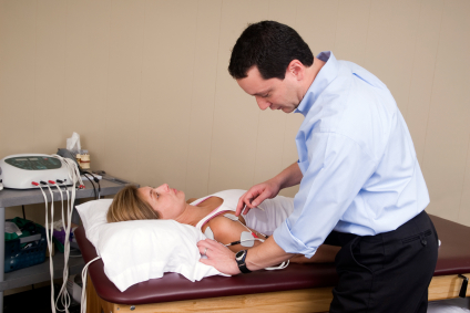
Examination

Inspection: The patient should be examined in sitting position with his upper torso and upper limbs exposed upto the waist.

Attitude: The position of the limb on inspection should be noted. In fractures of clavicle and anterior dislocation of shoulder, the patient often supports the injured limbs with the other hand. The arm segment may appear short or long depending on in fracture neck of scapula, there will be lengthing of the arm.

Swelling or deformity: In anterior dislocation of shoulder, the anterior axillary fold may be abnormally prominent due to the presence of head of humerus. A swelling along the line of clavicle, diffuse swelling surrounding the proximal humerus may be seen in fractures of the underlying bones. The lateral end of clavicle may appear to be prominent in acromioclavicular joint injuries. The medial end of clavicle may be seen prominently in sternoclavicular injuries.

Shoulder contour: Normally, the shoulder has a round contour due to prominence of the greater tuberosity beneath the deltoid muscle. The greater tuberosity projects beyond the edge of acromion process giving the normal contour. In dislocation of the shoulder joint, due to loss of projection of greater tuberosity, the normal contour will be lost. This is a valuable sign of dislocation. In deltoid paralysis due to axillary nerve injuries, there may be wasting of the muscle causing apparent loss of contour of the shoulder. The shoulder contour may be masked by diffuse swelling associated with fractures of the proximal humerus.

Bony arch: The bony arch is formed by the clavicle, acromion process and spine of scapula. Any deformity in the bony arch should be noted for.
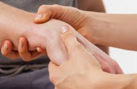
Palpation

The bony points to be palpated are: clavicle, proximal humerus, acromion process, spine and borders of scapula for signs of fracture.

Clavicle: By standing behind the sitting patient, the examiner places both his hands on the medial end of clavicle and runs his fingers along the shaft of both the clavicles. Any irregularity, gap or crepitus should be looked for. In acromioclavicular joint dislocation, the lateral end of clavicle may be displaced upward. On pressing the lateral end of clavicle, it depresses and bounces back like a piano key.

Proximal humerus: By standing on the side of the patient, the elbow is flexed and the proximal humerus is palpated bimanually by keeping one hand and the medial surface of arm and other on the outer surface of the arm. By standing behind the patient, the examiner slides his fingers down from the acromion process to the arm to palpate the greater tuberosity and proximal humerus. If the head of humerus is in normal position, then there will be a bony resistance to palpation. In dislocations, there will be an empty feeling in the shoulder region. The head may be palpable in either of axillary folds. Diffuse tenderness may be present in fractures of proximal humerus. In an intact humerus, the medial epicondyle will be in the same direction as that of the head of humerus.

Scapula: The acromion process and spine of scapula are palpated for irregularity, bony tenderness and crepitus. The axillary and vertebral borders are palpated for signs of fracture. The coracoids process is situated half an inch below the clavicle at its junction with medial two third and lateral one third. Fracture neck of scapula is diagnosed by axial pressure applied through the arm with the elbow flexed.

Movements

Both active and passive movements of the shoulder should be tested. In anterior dislocation of the shoulder, the patient will not be able to touch the opposite shoulder with his hand of affected extremity. This is called Dugas test.

Tests for detecting anterior dislocation of shoulder:

Hamilton ruler’s test: In normal persons, a straight ruler cannot be placed between the acromion process and lateral epicondyle because of the presence of greater tuberosity in its normal position. In dislocation of the shoulder, a ruler can be placed.

Callaway’s test: The vertical circumference of the axillary is increased in dislocation of shoulder due to the presence of head in the anterior axillary fold.

Measurements

The length of the arm is measured from the angle of acromion process to the lateral epicondyle.

Neurological examination: In fracture of the clavicle, brachial plexus may be injured. In fractures and dislocations of humorous, axillary nerve may get damaged. Axillary nerve damage may manifest as paralysis of deltoid muscle and anesthesia in skin over the lower part of deltoid muscle.

Active physical Therapy providing state-of-the-art physical therapy throughout the state of Maryland having multiple locations located in (Aspen Hill/ Layhill Road, Clinton, Clinton WHC, College Park/ Berwyn Heights, Columbia/ Elkridge, Columbia Aquatic, Gaithersburg/ Germantown, Hyattsville/ Langley Park, Landover, Laurel, Oxon Hill/ Temple Hills, Rockville), Washington, D.C (Washington D.C. N.W./ Near GWU , Washington D.C. N.E./ Brookland CUA, Washington D.C. S.E./ Capitol Hill), Western Maryland (Frederick, Hagerstown), Southern Maryland (California/ Lexington Park, Fort Washington La Plata, Prince Frederick, Waldorf), and Baltimore Metro area (Baltimore/ Mt. Vernon, Dundalk, Glen Burnie, Rosedale/ Near Franklin Sq. Med. Ctr.). We specialize in evaluation and treatment of acute and chronic conditions of the Upper Extremity. Experienced, Qualified and Skilled Certified therapists and our dynamic clinical staff focus on providing personalized attention, individual care, and a positive friendly environment during your treatment session. You can also make your appointment online to start your treatment within 24 to 48 hours at Active Physical Therapy, For more information just visit our Website:-http://active-physicaltherapy.com/

Treatment of Hand Joint Pain:

Volar Barton (Palmar rim dislocation) is a palmar rim fracture of distal radius. The most common cause of this type of fracture is a drop on an outstretched hand.

Mechanism

It is due to palmar tensile stress and dorsal shear stress and is usually combined with Radial styloid fracture.

Clinical Features

  • Pain

  • Swelling

  • Tenderness

  • Loss of wrist movements

  • Palmar flexion is grossly restricted and painful

Radiograph

Palmar rim of distal radial articular surface is displaced dorsally. Proximally and posteriorly and may be associated with dorsal subluxation of the wrist.

Treatment

Conservative

Reduction is simple, but retention is difficult. Long arm cast is used.

Surgery

If reduction does not remain satisfactorily with the wrist in neutral or slight palmar flexion, fixation with K-wire, external fixators and buttress plate, etc. may be done. Ellis T-’shaped buttress plate fixation is the preferred method of treatment.

Active Physical Therapy provides state of the art physical therapy throughout the state of Maryland.  Active’s friendly staff looks forward to assisting you in making your appointment in any of our clinics. Contact Us

http://www.active-physicaltherapy.com


Cubital Tunnel Syndrome: Causes, Symptoms and Treatment

Cubital Tunnel Syndrome is the second most common nerve compression, caused by increased pressure on the ulnar nerve at the elbow. The people, who repeatedly lean on their elbows or bend their elbows for sustained periods, are more likely to get affected by this condition. This condition is more common in men than women.

What do you mean by Cubital Tunnel Syndrome?

Cubital Tunnel Syndrome affects ulnar nerve where it crosses the inside edge of the elbow. This inside edge is relatively unprotected area and ulnar nerve here is entrapped between bone and skin in the tunnel called ‘Cubital Tunnel’.  When the ulnar nerve in the cubital tunnel gets compressed or irritated due to an injury or pressure, it called cubital tunnel syndrome. This condition affects the Baseball Pitchers more, as the twisting motion used to throw a slider can easily damage ligaments in their elbow.

What are the common causes of Cubital Tunnel Syndrome?

Cubital Tunnel Syndrome can occur due to following possible reasons:

  • Sustained bending of elbow during sleep
  • Leaning over your elbow for longer time
  • Fluid build-up in the elbow
  • A direct blow to inside edge of the elbow
  • Previous fracture, injury or dislocation of the elbow
  • Arthritis of elbow or bone spurs
  • Any cyst near the elbow
  • Repetitive bending, twisting or flexion of elbow during painting, playing an instrument or using power tools
  • Obesity or other health conditions like hypothyroidism or diabetes

What are the potential signs and symptoms of Cubital Tunnel Syndrome?

Typical symptoms of Cubital Tunnel Syndrome may include:

  • Numbness and tingling sensation or intermittent pain in the elbow
  • Pain in the elbow extending to your forearm and hand
  • Weakness in hand and fingers
  • Being unable to straighten your ring and little finger
  • Tenderness in the inside edge of the elbow
  • Difficulty in gripping and holding the objects
  • Reduced ability to pinch the thumb and little finger
  • Claw-like deformity of the hand

How can physical therapy help to treat Cubital Tunnel Syndrome?

Physical therapy treatment for Cubital Tunnel Syndrome includes patient education along with identification, modification and limiting the causes resulting into nerve inflammation. Apart from this, physical therapists may suggest:

  • Ergonomic considerations and changes in patterns of activities causing the condition
  • Specific elbow padding may be provided for work and leisure activities as well
  • Splinting at night may be suggested to limit range of motion enhancing the compression
  • Ulnar gliding exercises and modalities are practiced to relieve the painful symptoms
  • Modification of job activities, if needed, may be suggested
  • Therapists will suggest you different ways to rest your elbow and also educate you about the ways to use your elbow without putting any extra strain in it.
  • Heat or cold treatments may be applied to ease the pain
  • Strengthen and stretching exercises are used to strengthen the muscles of the forearm
  • Range-of-motion exercises are used to help you to return full length of muscles that are shortened due to protective posturing

Contact Active Physical Therapy for the state-of-art treatment of any of your musculoskeletal problems. Our certified ad dedicated physical therapists design individualized treatment plans depending upon your condition and problem.

Radial Tunnel Syndrome

What do we mean by Radial Tunnel Syndrome? What are its potential causes, symptoms and treatment options?

Radial Tunnel Syndrome 1Most of the athletes visit physical therapists for their lateral elbow pain. Physical therapists are of the view that Radial Tunnel Syndrome (RTS) occurs due to swelling or irritation of radial nerve because of the friction caused due to compression by muscles.

What do you mean by Radial Tunnel Syndrome?

Radial Tunnel Syndrome occurs when the radial nerve gets compressed where it passes through a tunnel near the elbow. This compression happens in proximal forearm where the radial nerve splits into PIN (main trunk) and sensory branch (minor trunk).

What are the causes of Radial Tunnel Syndrome (RTS)?

Pain of Radial Tunnel Syndrome occurs due to the pressure on radial nerve. Other causes of Radial Tunnel Syndrome include:

  • Injury
  • Bone Tumors
  • Inflammation/Swelling of surrounding tissues
  • Non-cancerous fatty-tumors
  • Too Small tunnel
  • Repetitive bending, gripping and pinching of wrist
  • Direct blow to the outside of elbow
  • Continuous twisting of arm

What are the potential symptoms of Radial Tunnel Syndrome (RTS)?

The symptoms of Radial Tunnel Syndrome start getting worse when you bend your wrist backward or hold an object with a stiff wrist. Following are the symptoms of Radial Tunnel Syndrome:

  • Tenderness and pain on the outside of elbow
  • Pain in the forearm and hand during activity
  • Minimized grip and forearm strength
  • Pain that worsens while rotating wrist
  • Cutting and stabbing pain at top of the forearm or at the back of hand, when you straighten your fingers
  • Numbness and tingling down the forearm

How can Physical Therapy be helpful to treat Radial Tunnel Syndrome (RTS)?

Fore mostly, the physical therapist will advise you to avoid the repetitive activities which involve your wrists. If Radial Tunnel Syndrome is caused due to your work site conditions, your work site conditions would be modified. Apart from these, following therapeutic techniques would be administered to treat the condition:

  • Stretching and strengthening exercises would be administered to reduce pain
  • Cold and heat therapies would be practiced
  • Range of motion exercises would be administered to restore tissue flexibility and normal functioning of the joints
  • You would be advised to wear night-splint to limit your elbow movements at night
  • Soft –tissue massage would be used to promote blood circulation
  • Modalities like ultrasound, moist heat and electrical stimulation may also be used
  • Spinal manual therapy may be used to improve neural mobility
  • Ergonomic interventions like; load management, postural awareness and workstation modification are used
  • Functional strengthening exercises are used to treat Radial Tunnel Syndrome

 Contact   Active Physical Therapy for the state-of-art treatment of your overuse injuries. Our certified and skilled physical therapists are always ready to help you with customized treatment plans as per your needs and requirements.

Sinus Tarsi Syndrome

Sinus Tarsi Syndrome

Sinus Tarsi Syndrome: Symptoms, Causes and Treatment

Sinus Tarsi Syndrome (STS) is a type of foot pathology, resulting either from the traumatic injury or recurrent injuries or sprain to the ankle during running or walking on a flat foot. It mostly hits athletes or dancers whose professions require a lot of jumping, sudden or quick movements and sudden stops.

 What do we mean by Sinus Tarsi Syndrome (STS)?

The word ‘sinus’ commonly refers to cavity in the bone. The cavity, Sinus Tarsi, is a small cylindrical cavity outside the ankle between the talus and calcaneous bones. Swelling around the Sinus Tarsi region or injury to any of the surrounding ligaments results in Sinus Tarsi Syndrome.

What are the common symptoms associated with Sinus Tarsi Syndrome?

Symptoms related with Sinus Tarsi Syndrome arise gradually over a period of time. Its symptoms are worse during morning but start improving as you warm up. Its symptoms include:

  • Sharp and pinching pain at the top and/or outer side of foot and ankle
  • Pain worsens during the activities like; walking or jumping
  • Stiffness in the ankle
  • Instability is felt while walking or running on uneven ground or slopes and during jumping or changing directions
  • Sinus Tarsi region may be felt tender when touched
  • Pain during ankle movements; especially when you move the sole of foot inwards or downwards
  • Pain intensifies with weight-bearing

What are the causes of Sinus Tarsi Syndrome?

The exact reason of Sinus Tarsi Syndrome is still a matter of debate. Trauma to the ankle is considered to be the most common cause of this pathological condition. Other than this, below mentioned factors give arise to Sinus Tarsi Syndrome:

  • An inversion injury to the foot that is not treated properly
  • Over growth of nerve or fat tissues in the cavity
  • Deviations in bone structures
  • Bone Spurs
  • Arthritis and Bone bridges
  • Ganglion Cysts
  • Foot Deformities (like Flat Foot)
  • Sitting with your feet tucked under you
  • Poor foot Bio-mechanics
  • Inappropriate Footwear
  • Poor flexibility and muscle weakness

How can Sinus Tarsi Syndrome be treated?

Physical Therapy treatment under the guidance and surveillance of expert Physical Therapist is the best treatment option available to treat Sinus Tarsi Syndrome. Therapeutic techniques employed by Physical Therapists not only speed up the recovery but also reduce the chances of recurrence. A complete Physical Therapy Treatment plan consists of:

  • Initially, RICE (Rest, Ice, Compression, Elevation) therapy is advised to to reduce and eliminate pain and to help tissues to heal
  • Activity triggering pain and swelling is identified and eliminated to reduce muscle tension
  • Stable shoes, an ankle sleeve or brace and over the counter or special orthotics are recommended
  • Electrotherapy, laser therapy and cryotherapy is employed

After the pain and swelling are subsided, physical therapists perform following exercises to enable the patient to return to normal activities:

  • Joint mobilisation exercises  are performed to reduce stiffness
  • Stretching exercises including; calf stretches and strengthening exercises are performed
  • Balance Training is provided to prevent  instability
  • Bio-mechanical correction is advised
  • Flexion exercises are administered to strengthen the muscles

 Contact Active Physical Therapy for the state-of-art and effective treatment of any of your musculoskeletal problem and disorders. Our proficient physical therapists create and develop customized treatment plans while taking into view your needs and urgencies.

Turf Toe

What do we mean by Turf Toe?

Turf Toe is the condition referring to the sprain of ligaments around the big toe joint. The injury is referred to as ‘Turf Toe’ as it generally affects the athletes who play on the artificial turf like; football, soccer or baseball players. It is also a common form of injury among the dancers.

But it does not imply that Turf Toe happens to athletes only; anyone whose big toe gets stubbed or jammed suddenly with enough force can develop this condition.

What are the causes leading to Turf Toe?

This injury occurs, when an athlete jams his toe forcefully into the ground or repeatedly pushes it off or bends it backward beyond the normal limits. Apart from these, another contributing factors to Turf Toe Include:

Turf Toe

  • Jamming of the foot on the ground, while rest of the body is pushed forward.
  • Wearing soft-soled foot wears, offering little or no protection to the fore foot.
  • Running and jumping on the hard artificial fields lead to the jamming of big toe and hence to the turf toe.
  • Repeated sprain over the ligaments with enough sudden force can lead to turf toe.
  • Body weight of the person
  • Direct injury that damages the bone under the cartilage.
  • Previous toe or fore foot injuries
  • Positioning of the body while playing sports
  • Yours ankle’s range of motion

What are the symptoms of Turf Toe?

The symptoms of Turf Toe include:

  • Pain at the base of the big toe
  • Tenderness in the ball of foot and big toe
  • Swelling of toe and ball of foot
  • Popping sound can be heard at the time of injury
  • Decreased mobility
  • Bruising of ball of foot and big toe
  • Inefficiency to put weight on the ball of injured foot
  • Failure to push off on the big toe

What are the treatment methods prescribed for Turf Toe?

The best way to treat Turf Toe is to keep the weight or pressure off the toe during the healing process. Treatment methods may include:

  • RICE (Rest, Ice, Compression and Elevation) therapy may be administered to reduce the swelling and pain in the affected area.
  • Athletes are advised to use orthotics or wear stiff soled shoes to secure the toe from more sprain or hyperextension.
  • Strapping or taping of the injured toe to the other toes to provide support and stability.
  • Doctors may immobilize the injured foot in a cast or special walking boot, in order to protect the affected joint

 After a period of immobility and rest, to re-establish the functioning of affected toe; physical therapists practice the following rehabilitation exercises:

  • Passive range of motion exercises are performed to regain function and mobility
  • Flexion and Extension exercises are performed
  • Strengthening exercises like; toe raises, calf raises, squats and lunges are performed
  • Conditioning and Agility exercises are performed

Contact Active Physical Therapy for the proficient treatment of any of your musculoskeletal problems or injuries. Our proficient and eminent physical therapists create customized treatment plans to help you to regain your health and agility.

Treatment for DeQuervain’s Tendonitis

What do you mean by DeQuervain’s Tendonitis?

DeQuervain’s Tendonitis is a painful condition that occurs when the tendons running from the back of your thumb down the side of your wrist get swollen and irritated. This swelling in turn, constricts the canal at the base of your wrist. So, every time when you turn your wrist to hold something or make a fist, it starts aching or hurting.

What are the causes of DeQuervain’s Tendonitis?

DeQuervain’s Tendonitis occurs when you continue with the activities which put strain on your tendons at back / outer aspect of thumb. Factors and activities listed below lead to DeQuervain’s Tendonitis:

de Quervain’s Syndrome.

  • Sports activities like; golf, ten pin bowling, rowing
  • Manual work like; painting, chopping wood, sewing, knitting, use of vibrating machinery
  • Constant lifting of children
  • Direct injury to wrist tendon
  • A fall or forceful movement
  • Injury to thumb, wrist elbow, neck, shoulder and upper back
  • Excessive training
  • Weakness of muscles
  • Muscle Stiffness
  • Tightening of joints
  • Faulty sporting technique
  • Bad posture
  • Poor ergonomic set up
  • Insufficient warm up

 What are the potential signs and symptoms of DeQuervain’s Tendonitis?

Following factors can be stated as potential symptoms of DeQuervain’s Tendonitis:

  • Ache or tightness at the back of thumb or wrist
  • Swollen tendon
  • Feeling of pain when tendon is touched
  • Occasional pins and needles feeling in the thumb
  • Weakness in the wrist, hand and thumb
  • Numbness in the Index finger
  • Movement of thumb and wrist is hindered due to pain and inflammation

How physical Therapy can help to treat DeQuervain’s Tendonitis?

DeQuervain’s Tendonitis is treated using medications, physical or occupational therapy. Discontinuing the activities that affect and worsen your condition, can also help a lot to cure and heal the ailment. Following techniques are employed by the physical therapists to treat DeQuervain’s Tendonitis:

  • Initially, the physical therapist will advice to rest your thumb and wrist to reduce inflammation. At times, splint and braces are used to keep them straight.
  • Manual hands- on therapy is used to treat DeQuervain’s Tendonitis.
  • Soft tissues massage, Trigger point releases, Joint mobilization therapies are administered to improve functioning and relieve strain on tendons.
  • Flexion, Extension and Rotation exercises are performed to strengthen the tendons.
  • Range of motion exercises are administered to loosen and mobilize the stiff tendon.
  • Strengthening exercises are performed to restore functioning and promote healing as well.
  • Stretches are administered to improve the flexibility of the affected tendon.

 At Active Physical Therapy, our eminent and skilled physical therapists are always ready to help you out with any of your musculoskeletal ailments and disorders. Your wellbeing is our top most priority. We are always ready to serve you with best of the treatment plans as per your needs and requirements.


Lumbar Stabilization Program

Lumbar Stabilization: Physical Therapy  Treatment & Management

Lumbar Stabilization is an exercise based program including variety of strengthening and flexibility exercises to heighten a person’s range of motion and as well to strengthen his muscles to support spine. This technique not only improvises his current physical conditions and symptoms but also acknowledge him about efficient movement and safe postures. Lumbar Stabilization program further helps you to find and maintain neutral spine position.

Role of a Physical Therapist

 Before planning and designing a well suited Lumbar Stabilization program, a physical therapist along with your referring physician; thoroughly examines and evaluates you, as the conditions of each individual are unique and specific. Thereafter a multi- dimensional program including strengthening, training, flexibility and endurance exercises is prescribed accordingly to the individual.

Lumbar Stabilization Program

Objectives or Goals of Lumbar Stabilization Program :

  • To attain a neutral spine position
  • To lower or lessen low back pain
  • To cure soft tissue injury like muscle strain
  • To lessen or eliminate lower back injuries that occur due to sudden jerks or movements
  • To exercise control over the forces acting on spine
  • To promote and heighten functional stability

Exercises involved in Lumbar Stabilization Program

During all the exercises performed under Lumbar stabilization program, a neutral spine position is attempted to attain and maintain. As the patient learns to maintain a neutral spine position during current exercise, he is progressed to the next exercise. All the exercises performed under Lumbar Stabilization are patient specific. Following exercises may be recommended under Lumbar Stabilization program :

  • Hamstring Stretch :This is a passive form of exercise, where little muscle effort is used or employed.
  • Pelvic Tilt : An active form of exercise making use of abdominal muscles to move the spine.
  • Arm or Leg raises : A dynamic exercise form, making use of arm or  leg movements while maintaining neutral spine position.
  • Stretching and aerobic exercises are also incorporated in Lumbar Stabilization physical therapy program.
  • Flexibility training is promoted to train muscles and to attain neutral position sine position.
  • Cardiovascular conditioning is combined with Lumbar Stabilization to promote total body strength and endurance.

At Active Physical Therapy, our professional and trained physical therapists ,plan and co-ordinate treatment modules which are designed according to your preferences, capabilities, needs and urgencies. Our treatment modules will not only heal your current ailment but also poses a check on the further ones.

Physical Therapy after a Hip Replacement

Hip Replacement: On the Road to Recovery

When the arthritis in the hip begins to deteriorate and damage the hip joint gradually, hip replacement surgery is performed. But only surgery does not make for the normal and successful functioning of the joint. The battle is won completely when with the help of physical therapy muscles around the hip joint along with the muscles of legs are strengthen and your implanted joint becomes mobilize and begins to work like a normal hip joint.

Hip Replacement Rehabilitation

Need of Hip Replacement Therapy

 Initially after surgery, patients feel stiffness and pain in the hip. So minor Physical Therapy begins on the first day after surgery and is recommended and needed to :

  • Ease and overcome the surgical pain
  • Improve hip’s range of motion
  • Strengthen the muscles around the hip joint
  • Strengthen the muscles of legs
  • Prevent clotting of blood
  • Provide patient education regarding fall, preventions and walking without limp
  • Prevent the loosening and dislocation of the implant

Patient education provided during Hip Replacement Therapy:

  • Patients are advised not to pressurize and stress the hip joint with heavy lifting
  • Specific body posturing and sitting techniques are instructed
  • Patients are briefed not to cross the operated leg over the other one as it can cause the dislocation of the replaced joint
  • Patients are acknowledged about the precautions and safety measures that they should follow while performing the routine activities
  • Patients are educated about the available adaptive  and supportive equipment and their usage

 Exercises recommended under Hip Replacement Therapy:

Physical therapists suggest that to accomplish full recovery and to restore normal hip motion, regular exercises should be performed. During initial days, therapists recommend to exercise 20 to 30 minutes for 2-3 times a day. Following exercises may be suggested and administered by the physical therapists  :

  • Ankle Pumps: It involves pushing your foot up and down.
  • Ankle Rotations: It involves moving your ankle inward and outward the other foot.
  • Buttock Contractions: Involves tightening of buttock muscles.
  • Hip Extension: Involves contracting your hip muscles without holding your breath.
  • Hip Abduction and Adduction: Involves sliding of each leg in and out then back with straight knees and pointed toes.
  • Bridging: Includes lifting up the buttock with bent knees and flat feet.
  • Standing Exercises: These exercises are administered to help you to regain strength for standing independently.
  • Walking, Stair climbing and Descending Exercises: These exercises improvise  strength, mobility and endurance of your knees, hip and legs. Further more, these exercises also help you to regain your balancing skills.

Contact Active Physical Therapy if you are looking forward to the treatment plans that are designed according to your urgencies, preferences and capabilities. Our trained and proficient therapists will provide you with best possible assistance and care and help you to regain your health and well being.

How to cure Lumbar Spondylolysis?

Causes and Symptoms of Spondylolysis:

Lumbar SpondylolysisSpondylolysis happens when a crack forms in the bony ring on the back of the spinal column. Most commonly, this occurs in the low back. In this condition, the bone that protects the spinal cord fractures as a result of excessive or repeated strain. The area affected is called the pars interarticularis, so doctors sometimes refer to this condition as a pars defect.

This condition appears in six percent of children. It mainly affects young athletes who participate in sports in which the spine is repeatedly bent backwards, such as gymnastics, football, and karate.

Anatomy

The human spine is made up of 24 spinal bones, called vertebrae. Vertebrae are stacked on top of one another to create the spinal column. The spinal column gives the body its form. It is the body’s main upright support. The section of spine in the lower back is called the lumbar spine.

Each vertebra is formed by a round block of bone, called a vertebral body. A circle of bone attaches to the back of the vertebra. When the vertebrae are stacked on top of each other, these bony rings create a hollow tube. This tube, called the spinal canal, surrounds the spinal cord as it passes through the spine. Just as the skull protects the brain, the bones of the spinal column protect the spinal cord.

Causes of Lumbar Spondylolysis

Spondylolysis is thought to be caused by repeated strains that damage the lower spine over time. The repeated strains can eventually lead to an overuse injury in the pars interarticularis. The most common location for this to occur is in the lowest vertebra of the spine. This vertebra connects the spine to the pelvis. However, a problem with the pars can occur in any lumbar vertebra. It rarely happens in more than one vertebra at a time.

  • The vertebra initially responds to the abnormal strain by adding new bone cells around the injured area.
  • But if the injuries happen faster than the body can keep up with needed repairs, a crack may form in the weakened bone. This is called a stress fracture.
  • This type of fracture occurs in the pars, the area of bony ring between the pedicle and lamina.
  • The crack may affect only one side of the bony ring. However, it is equally common for the defect to occur on both sides.
  • When this happens, the vertebra is no longer held firmly in place by the facet joints on the back of the ring.
  • As a result, the vertebra is free to slip forward over the one below.
  • This slippage, which is closely related to spondylolysis, is called spondylolisthesis.

Symptoms

  1. People with spondylolysis may feel pain and stiffness in the center of the low back.
  2. Bending fully backward increases pain.
  3. Symptoms typically get worse with activity and go away with rest.
  4. Doctors refer to this type of back pain as mechanical pain because it most likely comes from excess movement between the vertebrae.
  5. Individuals may eventually experience pain that radiates down one or both legs.
  6. This pain may come from pressure and irritation on the nerves that exit the spinal canal near the fracture.
  7. When nerve pressure in the low back causes leg pain, doctors refer it as neurogenic pain.

Treatment

  • Recovery from this condition is much like nonsurgical treatment mentioned earlier.
  • Once you have rested your back to allow it to heal, your doctor may recommend that you work with a physical therapist a few times each week for four to six weeks.
  • In severe cases, patients may need a few additional weeks of physical therapy.

Active’s friendly staff looks forward to assisting you in making your appointment in any of our clinics. Please do not hesitate to contact us, we are dedicated to providing the best physical therapy service in Maryland.